Thursday, October 31, 2013

New dark matter detector sends first data from gold mine 1.5km underground

Scientists testing the Large Underground Xenon (LUX) experiment have reported promising scientific and technological results today.

They have set up the experiment to identify the nature of dark matter, an invisible substance that physicists believe is all around us, making up most of the matter in the universe, but that barely has any effect on our every-day lives.

Scientists have published the first results from the Sanford laboratory today, which, they say, validate the experiment's design and performance. The research challenges previous studies that claim 'sightings' of dark matter.
They are now beginning a process to uncover the exact identity of the dark matter particle - a process equivalent to the work done by the Large Hadron Collider in identifying the Higgs boson.

Seventeen universities and research institutes in the USA and Portugal, and Imperial College London, UCL, University of Edinburgh in the UK, run the LUX experiment, with most funding coming from the National Science Foundation and Department of Energy in the USA.

The new laboratory is sited in a former gold mine nearly one and a half kilometres below the Black Hills in the American state of South Dakota. Work on LUX started in 2008 and the experiment was completed ready for an initial run earlier in 2013.

Here, scientists are operating some of the world's most sensitive equipment in an extremely sheltered environment, because they are looking for tiny and extremely rare flashes of light that would indicate a collision between a dark matter particle and a normal matter particle.

"These instruments take many years to build and we are always pushing new technologies to the limit," says Dr Henrique Araújo from the Department of Physics at Imperial College London who leads the College's team working on LUX.

"It is very significant that LUX worked as designed when we finally pushed the 'on' button. Many experiments never reach this stage."

Physicists believe that dark matter accounts for about a quarter of the energy in the Universe, compared with ordinary matter, which makes up only a twentieth. The rest consists of the even more mysterious dark energy.

Since the experiment was installed underground in February, they have been looking for Weakly Interacting Massive Particles (WIMPs), which are the prime candidates to constitute the dark matter in our galaxy and in the Universe.

These particles are thought to have mass like normal particles and create a tiny gravitational pull, but cannot be observed directly since they neither emit nor absorb light at any wavelength. On the largest scales, its presence can be inferred from the motion of stars within galaxies, and of individual galaxies in galactic clusters.

Collisions between WIMPs and normal matter are rare and extremely difficult to detect because cosmic-ray particles from space can overwhelm the already faint flashes expected from WIMPs.

Few cosmic rays can penetrate as deep underground as the LUX experiment and the detector is further protected from background radiation by being immersed in a shielding tank of ultra-pure water.
"We are able to detect the faint flashes of light very effectively using good internal reflector materials and very sensitive photon sensors," says Dr Araújo.

"LUX has significantly higher sensitivity than the previous world's best dark matter experiments – especially for the lightest WIMPs, which cause the faintest signals."

The new LUX result challenges evidence from other experiments, such as CoGeNT and DAMA, where scientists have previously claimed to have data about the nature of WIMPs.

Dr Araújo says: "A number of previous results make it look like WIMPs exist with a particularly low mass. While this may still turn out to be the case, our new data reveal that, on that occasion, it was a case of mistaken identity."

HUNTING FOR DARK MATTER

A decade ago, scientists of the UK-led ZEPLIN programme deployed the first dark matter detector of this type underground at the Boulby mine in North Yorkshire.

"We had a pioneering role in what has become the world's most sensitive dark matter search technology, building and operating three detectors at Boulby."

"The last and most sensitive, ZEPLIN-III, was built by our team at Imperial – until we concluded our programme in 2011 – joining LUX soon after."

Scientists including Dr Araújo are already designing – and soon will start building – the next-generation experiment, LZ, which is the coming together of the two programmes – LUX and ZEPLIN.

With a 7-tonne liquid xenon target, LZ will be 30 times larger than LUX and have over 100 times better range. It will be so sensitive that it will be limited only by the interference of background signals from astrophysical neutrinos. These similarly illusive particles were once a candidate to explain the dark matter problem — but physicists now know they are not massive enough to do the job.

LZ is a collaboration of 26 institutes in the US, UK, Portugal and Russia. Dr Araújo from Imperial leads the UK team on LZ, which counts also with colleagues from Edinburgh, UCL, Oxford and Sheffield universities as well as the Rutherford Appleton and Daresbury national laboratories.

HOW DOES IT WORK

At the heart of the experiment is a 6-foot-tall titanium 'thermal flask' filled with almost a third of a tonne of liquid xenon, cooled to minus 100 degrees centigrade.

When a WIMP hits a xenon atom it recoils – like a white billiard ball striking the opening triangle of coloured balls in a game of snooker – and photons of light are emitted; at the same time, this interaction also releases electrons from surrounding atoms.

The electrons are drawn upward by an electrical field and get absorbed into a thin layer of xenon gas at the top of the tank, releasing more photons.

Light detectors in the top and bottom of the tank are each capable of detecting these two photon signatures. The locations of the two signals can be pinpointed to within a few millimetres.


The energy of the interaction can be precisely measured from the brightness of the pulses of light. Any particles interacting in the xenon will cause these signals, but WIMP interactions are expected to have characteristic sizes which are quite different from those caused by ordinary particles.

Source: http://www3.imperial.ac.uk/newsandeventspggrp/imperialcollege/newssummary/news_30-10-2013-15-31-20

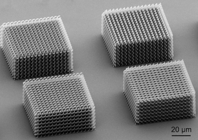
3D Photonics

After the invention of the laser in 1960, optics turned into photonics. In todays telecommunication technology, photons have already become the main carrier of information. During the last decades, a number of pioneering developments such as, e.g., the concepts of photonic crystals and metamaterials have opened the door to a completely new class of materials. Molding the flow of light as well as controlling the dynamics of photons are two key issues. The properties of such nanostructured materials are subject of current research activities.


Shown below is a series of structures fabricated by means of the direct laser writing process achieved with Photonic Professional systems.


For more info: http://www.nanoscribe.de

3D square spiral structure out of SU-8.


Three-dimensional photonic crystal of cubic symmetry fabricated
by two photon polymerization.


Three-dimensional photonic crystal of cubic symmetry fabricated
by two photon polymerization.


Bi-chiral photonic crystals, produced by the group of
Prof. Dr. Martin Wegener (KIT)


3D photonic quasicrystal with a five-fold rotational symmetry.
The structure was written into the photoresist SU-8.
Image: Dr. Alexandra Ledermann (KIT)


Circular spirals seen from above. This structure was written into the
chalcogenide glass As2S3.


Further examples of 3D structures consisting of circular spirals.

3D photonic crystal structure according to the layout provided
by Mr. Ivan Shishkin, ITMO, Russia.


3D photonic crystalline diamond (PCD) structure according to
Physical Review Letters 100, 013901 (2008) and Physical Review
B 82, 115116 (2010). With friendly permission of
Prof. Dr. Keiichi Edagawa, Institute of Industrial Science,
The University of Tokyo. Material: IP-L 780.


Light cone array along the publication "Microphotonic
parabolic light directors fabricated by two-photon lithography" by J. H.
Atwater, P. Spinelli, E. Kosten, J. Parsons, and C. Van Lare in Appl.
Physics Letters 99, 151113 (2011).


3D photonic crystal known under the acronym SP2 - standing
for slanted pore structure. Originally proposed for anisotropic
etching techniques or GLAD "2" stands for the number of separate
drilling/etching/GLAD-processes. With our technique these
structure can directly be written into a photosensitive material in one
step. Later on these structures can be replicated or inverted e.g. in
silicon with our techniques.


Plasmonic crystal alters to match light-frequency source

Scientists actively control strongly coupled plasmonic resonators.
Image credit: Gregory C. Dyer, Sandia National Laboratory
Gems are known for the beauty of the light that passes through them. But it is the fixed atomic arrangements of these crystals that determine which light frequencies are permitted passage.
Now a Sandia-led team has created a plasmonic, or plasma-containing, crystal that is tunable. The effect is achieved by adjusting a voltage applied to the plasma, making the crystal agile in transmitting terahertz light at varying frequencies. This could increase the bandwidth of high-speed communication networks and generally enhance high-speed electronics.
“Our experiment is more than a curiosity precisely because our plasma resonances are widely tunable,” says Sandia researcher Greg Dyer, co-primary investigator of a recently published online paper in Nature Photonics, expected to appear in print in that journal in November. “Usually, electromagnetically induced transparencies in more widely known systems like atomic gases, photonic crystals and metamaterials require tuning a laser’s frequencies to match a physical system. Here, we tune our system to match the radiation source. It’s inverting the problem, in a sense.”
The plasmonic crystal method could be used to shrink the size of photonic crystals, which are artificially built to allow transmission of specific wavelengths, and to develop tunable metamaterials, which require micron- or nano-sized bumps to tailor interactions between manmade structures and light. The plasmonic crystal, with its ability to direct light like a photonic crystal, along with its sub-wavelength, metamaterial-like size, in effect hybridizes the two concepts.
The crystal’s electron plasma forms naturally at the interface of semiconductors with different band gaps. It sloshes between their atomically smooth boundaries that, when properly aligned, form a crystal. Patterned metal electrodes allow its properties to be reconfigured, altering its light transmission range. In addition, defects intentionally mixed into the electron fluid allow light to be transmitted where the crystal is normally opaque.
Greg Dyer Plasmonic Chip
Sandia National Laboratories researcher Greg Dyer
investigates a tunable crystal that could increase
 the bandwidth of high-speed communication
 networks.
(Photo by Randy Montoya)
However, this crystal won’t be coveted for the beauty of its light. The crystal transmits in the terahertz spectrum, a frequency range invisible to the human eye. Scientists also must adjust the crystal’s two-dimensional electron gas to electronically vary its output frequencies, something casual crystal buyers probably won’t be able to do.
Following online release, the paper titled, “Induced transparency by coupling of Tamm and defect states in tunable terahertz plasmonic crystals,” is slated to appear in the November print edition ofNature Photonics.
In addition to Dyer, other authors are co-principal investigator Eric Shaner, with Albert D. Grine, Don Bethke and John L. Reno, all from Sandia; Gregory R. Aizin of The City University of New York; and S. James Allen of the Institute for Terahertz Science and Technology at the University of California, Santa Barbara.
The work was supported by the Department of Energy’s Office of Basic Energy Sciences (BES) and performed in part at the Center for Integrated Nanotechnologies (CINT), a Sandia/Los Alamos national laboratories user facility that is one of the five DOE BES Nanoscale Science Research Centers that are premier national user facilities for interdisciplinary research at the nanoscale. Together the NSRCs comprise a suite of complementary facilities that provide researchers with state-of-the-art capabilities to fabricate, process, characterize and model nanoscale materials, and constitute the largest infrastructure investment of the National Nanotechnology Initiative. The NSRCs are located at DOE’s Argonne, Brookhaven, Lawrence Berkeley, Oak Ridge, Sandia and Los Alamos National Laboratories. For more information about the DOE NSRCs, click here.

Sandia National Laboratories is a multi-program laboratory operated by Sandia Corporation, a wholly owned subsidiary of Lockheed Martin company, for the U.S. Department of Energy’s National Nuclear Security Administration. With main facilities in Albuquerque, N.M., and Livermore, Calif., Sandia has major R&D responsibilities in national security, energy and environmental technologies and economic competitiveness.
Sandia news media contact: Neal Singer, nsinger@sandia.gov, (505) 845-7078

Giant atom eats quantum gas

A team of experimental and theoretical physicists from the University of Stuttgart studied a single micrometer sized atom. This atom contains tens of thousands of normal atoms in its electron orbital. These results have been published in the renowned journal Nature.
Illustration of the system investigated: A highly excited Rydberg-atom, consisting of a single electron (blue), traveling on a large orbit far from the positively charged core (red). The Rydberg atom has the same spatial extent as the ultracold atomic cloud. The single electron is exciting oscillations, so called phonons, in the surrounding quantum gas.
The interaction of electrons and matter is fundamental to material properties such as electrical conductivity. Electrons are scattering from atoms of the surrounding matter and can excite lattice oscillations, so called phonons, thereby transferring energy to the environment. The electron is therefore slowed which causes electrical resistance. However, in certain materials phonons can surprisingly cause the opposite effect, so called superconductivity, where the electrical resistance drops to zero. Understanding the interaction of electrons and matter is therefore important goal in order to both answer fundamental questions as well as to solve technical problems.
A single electron is best suited for systematic investigations of such processes. For the first time, physicists from Stuttgart have now realized a model system in the laboratory, where the interaction of a single electron with many atoms inside its orbital can be studied. These atoms are from an ultracold cloud near absolute zero, a so called Bose-Einstein condensate.
The basic idea now is simple: Instead of using a technically challenging electron trap, the scientists are using the fact that in nature electrons are bound to a positively charged atomic core. In a classical picture, they are travelling on ellipsoidal orbits around the core. These orbits are usually very small, typically in the range below one nanometer. In order to achieve an interaction between an electron and many atoms, an atom is excited from a cloud consisting of 100.000 atoms using laser light. The orbit of a single electron then expands to several micrometers and a Rydberg atom is formed. On atomic length scales, this atom is huge, larger than most bacteria, which are consisting each of several billions to trillions of atoms. The Rydberg atom is then containing tens of thousands of atoms from the cold cloud. Thus, a situation is realized where the electron is trapped in a defined volume and at the same time interacts with a large number of atoms. This interaction is so strong that the whole atomic cloud, consisting of 100,000 atoms is considerably influenced by the single electron. Depending on its quantum state the electron excites phonons in the atomic cloud, which can be measured as collective oscillations of the whole cloud culminating in a loss of atoms from the trap.
The experimental observations in the group of Prof. Tilman Pfau could so far largely be explained by collaborative work with the theory group of Prof. Hans Peter Büchler. However, this work is only the basis for a series of further exciting experiments. According to the previous studies an electron is leaving a clear trace in the surrounding atomic cloud. Therefore imaging a single electron in a well defined quantum state seems to be feasible. Due to the impact on various fields, including quantum optics, these results were published in the highly respected journal Nature *).
This work has been realized within Sonderforschungsbereich SFB/TRR 21 (Control of quantum correlations in tailored matter) and has been supported by the Detusche Forschungsgemeinschaft DFG and the European Research Council.
*) J.B. Balewski, A.T. Krupp, A. Gaj, D. Peter, H.P. Büchler, R. Löw, S. Hofferberth and T. Pfau, Coupling a single electron to a Bose-Einstein condensate; Nature, doi:10.1038/nature12592

Further information: 
Prof. Tilman Pfau, Jonathan Balewski, 5. Physikalisches Institut, Tel. +49 711/685-64820, e-mail: t.pfau@physik.uni-stuttgart.de, j.balewski@physik.uni-stuttgart.de 
www.pi5.uni-stuttgart.de 

Incurable brain cancer gene is silenced

Gene regulation technology increases survival rates in mice with glioblastoma

Glioblastoma multiforme (GBM), the brain cancer that killed Sen. Edward Kennedy and kills approximately 13,000 Americans a year, is aggressive and incurable. Now a Northwestern University research team is the first to demonstrate delivery of a drug that turns off a critical gene in this complex cancer, increasing survival rates significantly in animals with the deadly disease.

The novel therapeutic, which is based on nanotechnology, is small and nimble enough to cross the blood-brain barrier and get to where it is needed -- the brain tumor. Designed to target a specific cancer-causing gene in cells, the drug simply flips the switch of the troublesome oncogene to "off," silencing the gene. This knocks out the proteins that keep cancer cells immortal.

In a study of mice, the nontoxic drug was delivered by intravenous injection. In animals with GBM, the survival rate increased nearly 20 percent, and tumor size was reduced three to four fold, as compared to the control group. The results will be published Oct. 30 in Science Translational Medicine.

"This is a beautiful marriage of a new technology with the genes of a terrible disease," said Chad A. Mirkin, a nanomedicine expert and a senior co-author of the study. "Using highly adaptable spherical nucleic acids, we specifically targeted a gene associated with GBM and turned it off in vivo. This proof-of-concept further establishes a broad platform for treating a wide range of diseases, from lung and colon cancers to rheumatoid arthritis and psoriasis."
Mirkin is the George B. Rathmann Professor of Chemistry in the Weinberg College of Arts and Sciences and professor of medicine, chemical and biological engineering, biomedical engineering and materials science and engineering.

Glioblastoma expert Alexander H. Stegh came to Northwestern University in 2009, attracted by the University's reputation for interdisciplinary research, and within weeks was paired up with Mirkin to tackle the difficult problem of developing better treatments for glioblastoma.
Help is critical for patients with GBM: The median survival rate is 14 to 16 months, and approximately 16,000 new cases are reported in the U.S. every year.

In their research partnership, Mirkin had the perfect tool to tackle the deadly cancer: spherical nucleic acids (SNAs), new globular forms of DNA and RNA, which he had invented at Northwestern in 1996, and which are nontoxic to humans. The nucleic acid sequence is designed to match the target gene.

And Stegh had the gene: In 2007, he and colleagues identified the gene Bcl2Like12 as one that is overexpressed in glioblastoma tumors and related to glioblastoma's resistance to conventional therapies.

"My research group is working to uncover the secrets of cancer and, more importantly, how to stop it," said Stegh, a senior co-author of the study. "Glioblastoma is a very challenging cancer, and most chemo-therapeutic drugs fail in the clinic. The beauty of the gene we silenced in this study is that it plays many different roles in therapy resistance. Taking the gene out of the picture should allow conventional therapies to be more effective."
Stegh is an assistant professor in the Ken and Ruth Davee Department of Neurology at the Northwestern University Feinberg School of Medicine and an investigator in the Northwestern Brain Tumor Institute.

The power of gene regulation technology is that a disease with a genetic basis can be attacked and treated if scientists have the right tools. Thanks to the Human Genome Project and genomics research over the last two decades, there is an enormous number of genetic targets; having the right therapeutic agents and delivery materials has been the challenge.

"The RNA interfering-based SNAs are a completely novel approach in thinking about cancer therapy," Stegh said. "One of the problems is that we have large lists of genes that are somehow disregulated in glioblastoma, but we have absolutely no way of targeting all of them using standard pharmacological approaches. That's where we think nanomaterials can play a fundamental role in allowing us to implement the concept of personalized medicine in cancer therapy."

Stegh and Mirkin's drug for GBM is specially designed to target the Bcl2Like12 gene in cancer cells. Key is the nanostructure's spherical shape and nucleic acid density. Normal (linear) nucleic acids cannot get into cells, but these spherical nucleic acids can. Small interfering RNA (siRNA) surrounds a gold nanoparticle like a shell; the nucleic acids are highly oriented, densely packed and form a tiny sphere. (The gold nanoparticle core is only 13 nanometers in diameter.) The RNA's sequence is programmed to silence the disease-causing gene.

"The problems posed by glioblastoma and many other diseases are simply too big for one research group to handle," said Mirkin, who also is the director of Northwestern's International Institute for Nanotechnology. "This work highlights the power of scientists and engineers from different fields coming together to address a difficult medical issue."

Mirkin first developed the nanostructure platform used in this study in 1996 at Northwestern, and the technology now is the basis of powerful commercialized and FDA-cleared medical diagnostic tools. This new development, however, is the first realization that the nanostructures injected into an animal naturally find their target in the brain and can deliver an effective payload of therapeutics.

The next step for the therapeutic will be to test it in clinical trials.

The nanostructures used in this study were developed in Mirkin's lab on the Evanston campus and then used in cell and animal studies in Stegh's lab on the Chicago campus.


###


Northwestern has one of nine Centers of Cancer Nanotechnology Excellence (CCNE) funded by the National Cancer Institute. Mirkin and Stegh are members of both the CCNE and the Robert H. Lurie Comprehensive Cancer Center of Northwestern University.

The title of the paper is "Spherical Nucleic Acid Nanoparticle Conjugates as an RNAi-Based Therapy for Glioblastoma."

The co-first authors are Samuel A. Jensen, Emily S. Day and Caroline H. Ko. In addition to these three, Mirkin and Stegh, other authors of the paper are Lisa A. Hurley, Janina P. Luciano, Fotini M. Kouri, Timothy J. Merkel, Andrea J. Luthi, Pinal C. Patel, Joshua I. Cutler, Weston L. Daniel, Alexander W. Scott, Matthew W. Rotz, Thomas J. Meade and David A. Giljohann, all from Northwestern.

Source: http://www.northwestern.edu/

Wednesday, October 30, 2013

Nanostructured metal oxide films formed using microplasma-assisted, reactive chemical vapor deposition


Michael J. Gordon, PhDUniversity of California (Santa Barbara) is investigating plasma-based routes for direct synthesis of nanoparticles and hierarchically-ordered/structured thin films and nanostructures which have useful optical, electronic, and catalytic properties. In particular, we have developed a hydrodynamically-stabilized, microplasma jet-based growth technique to realize a variety of metal oxide nanowires (e.g., CuO, PdO, NiO, Fe2O3, SnO2) on different substrates (e.g., Si and ITO) at high pressures (10-100 torr). See Fig. 3. Although many examples of nanowire growth using the vapor-liquid-solid (VLS) method with a catalyst particle exist, our work demonstrates that anisotropic growth can be realized without a catalyst, mask, or surfactant using microplasmas to create a directed, tunable flux of atoms, metastables, and clusters (i.e., by controlling ballistic vs. diffusional aggregation phenomena) for anisotropic growth. Variants of the microplasma technique are currently being used to synthesize porous and textured metal and alloy films as well as nano- and microstructured oxides for electrocatalysis, gas sensing, and solar cells.

Read more: http://acswebcontent.acs.org/prfar/2012/Paper12047.html

Four-Color Nanoprobe Could Increase Accuracy Of Cancer Detection

In Living Color
A four-color probe for messenger RNA biomarkers consists of a gold nanoparticle (yellow circle) decorated with short loops of DNA (blue), each with a fluorescent dye on the end (colored circles). When the DNA loops are closed, and the fluorophores are near the gold nanoparticles, the gold quenches the fluorescence (left). But when the DNA sequences bind to their target mRNAs, they open, and the dyes can glow (right).
Credit: Anal. Chem.


Medical Diagnostics: Gold nanoparticles armed with fluorescent molecular beacons can recognize four cancer biomarkers at the same time, reducing the chance of a false

Cancer researchers have long looked for ways to detect cancerous cells at early stages of the disease. Tools that help doctors diagnose cancer early could increase the success of treatments. One approach has been to search for specific biomarkers, such as RNA molecules, that cancer cells produce large amounts of. But because healthy cells also produce these molecules, testing for just one or two such biomarkers runs the risk of getting a false positive result. Now, researchers in China have developed a cancer probe designed to reduce the chance of falsely identifying cancer cells. They have synthesized a four-color fluorescent probe that can detect the presence of four RNA biomarkers at the same time (Anal. Chem. 2013, DOI:10.1021/ac402700s).
In 2007, researchers at Northwestern University, led by Chad A. Mirkin, developed a strategy to spot these biomarkers by modifying gold nanoparticles to release “nanoflares”—short strands of DNA with a fluorescent dye attached—in the presence of a target messenger RNA sequence (J. Am. Chem. Soc. 2007, DOI: 10.1021/ja0776529). Last year, a team led by Bo Tang ofShandong Normal University, in China, built on that idea, creating a fluorescent three-color probe that used nanoflares to detect three different mRNA biomarkers simultaneously in live cells (Angew. Chem. Int. Ed. 2012, DOI:10.1002/anie.201203767).
But three colors were not enough—an extra color would help to further prevent false positive results, says Shandong Normal University’s Na Li, co-lead author on the new research. Also, Tang and colleagues wanted to cut down the time it took to spot the biomarkers. To meet these two goals, Tang’s team developed a four-color probe with a different signaling mechanism: Instead of using nanoflares, they used so-called molecular beacons that glowed in the presence of mRNA biomarkers.
To make the molecular beacons, the researchers started with snippets of single-stranded DNA that bind to one of four mRNA biomarkers from breast and liver cancer cells. They attached a different fluorescent dye to the end of each of the four DNA strands and then coated 13-nm-diameter gold nanoparticles with the labeled DNA. The strands fold onto themselves to form loops anchored at one end to the nanoparticle. When each fluorescently labeled loop is wrapped up tight, the fluorescent dye rests close to the gold nanoparticle, and the gold quenches the fluorescence. But when the DNA loops bind to their target mRNA sequences, they unfold. That opening moves the fluorescent dyes perched at the loose ends of the DNA strands away from the gold nanoparticle, allowing them to shine.
To test their nanoprobes, the team incubated the armored gold nanoparticles with breast and liver cells, both cancerous and normal ones. The cells take up the particles, and the probes bump into mRNAs inside the cytoplasm. The researchers then measured the fluorescence produced in the cells using a confocal microscope. Both types of cancer cells lit up with all four colors, indicating that all the tested biomarkers were being overexpressed. The healthy breast cells remained dark. But the healthy liver cells glowed in two colors, showing the importance of looking for multiple mRNAs to distinguish between healthy and cancer cells, Li says.
The process of unfolding the DNA loops is faster than sending out a nanoflare, so the new probe gets answers from living cells within three hours, instead of 12 hours with the nanoflare probes, Li says.
The new work “represents a major advancement in the design of gold-nanoparticle-based nanobeacons,” says Chunhai Fanof the Shanghai Institute of Applied Physics, whose group also has made three-color fluorescent nanoprobes. He explains that the surface chemistry necessary for the team to add a fourth color to the nanoprobe is tricky: Adding a new color means determining how to deposit the proper ratio of the probes and picking the dyes to prevent an overlap of colors from each beacon.

Tuesday, October 29, 2013

Diagnostic Devices the Size of a Credit Card Are Now a Possibility

A microfluidic bioreactors consists of two chambers separated by
a nanoporous silicon membrane. It allows for flow-based assays
using minimal amounts of reagent. The ultra-thin silicon membrane
provides an excellent mimic of biological barrier properties.
NOTE: This image combines two exposures in order to capture the
brighter and darker parts of the scene, which exceed the dynamic
range of the camera sensor. The resulting composite is truer to what the
eye actually sees.
The ability to shrink laboratory-scale processes to automated chip-sized systems would revolutionize biotechnology and medicine. 

For example, inexpensive and highly portable devices that process blood samples to detect biological agents such as anthrax are needed by the U.S. military and for homeland security efforts. One of the challenges of "lab-on-a-chip" technology is the need for miniaturized pumps to move solutions through micro-channels. Electroosmotic pumps (EOPs), devices in which fluids appear to magically move through porous media in the presence of an electric field, are ideal because they can be readily miniaturized. EOPs however, require bulky, external power sources, which defeats the concept of portability. But a super-thin silicon membrane developed at the University of Rochester could now make it possible to drastically shrink the power source, paving the way for diagnostic devices the size of a credit card.

"Up until now, electroosmotic pumps have had to operate at a very high voltage—about 10 kilovolts," said James McGrath, associate professor of biomedical engineering. "Our device works in the range of one-quarter of a volt, which means it can be integrated into devices and powered with small batteries."

McGrath's research paper is being published this week by the journal Proceedings of the National Academy of Sciences.

McGrath and his team use porous nanocrystalline silicon (pnc-Si) membranes that are microscopically thin—it takes more than one thousand stacked on top of each other to equal the width of a human hair. And that's what allows for a low-voltage system.

A porous membrane needs to be placed between two electrodes in order to create what's known as electroosmotic flow, which occurs when an electric field interacts with ions on a charged surface, causing fluids to move through channels. The membranes previously used in EOPs have resulted in a significant voltage drop between the electrodes, forcing engineers to begin with bulky, high-voltage power sources. The thin pnc Si membranes allow the electrodes to be placed much closer to each other, creating a much stronger electric field with a much smaller drop in voltage. As a result, a smaller power source is needed.

"Up until now, not everything associated with miniature pumps was miniaturized," said McGrath. "Our device opens the door for a tremendous number of applications."

Along with medical applications, it's been suggested that EOPs could be used to cool electronic devices. As electronic devices get smaller, components are packed more tightly, making it easier for the devices to overheat. With miniature power supplies, it may be possible to use EOPs to help cool laptops and other portable electronic devices.

McGrath said there's one other benefit to the silicon membranes. "Due to scalable fabrication methods, the nanocrystalline silicon membranes are inexpensive to make and can be easily integrated on silicon or silica-based microfluid chips."

Monday, October 28, 2013

UNC neuroscientists discover new 'mini-neural computer' in the brain

This is a dendrite, the branch-like structure of a single
neuron in the brain. The bright object from the top is a
pipette attached to a dendrite in the brain...
Dendrites, the branch-like projections of neurons, were once thought to be passive wiring in the brain. But now researchers at the University of North Carolina at Chapel Hill have shown that these dendrites do more than relay information from one neuron to the next. They actively process information, multiplying the brain's computing power.
"Suddenly, it's as if the processing power of the brain is much greater than we had originally thought," said Spencer Smith, PhD, an assistant professor in the UNC School of Medicine.

His team's findings, published October 27 in the journal Nature, could change the way scientists think about long-standing scientific models of how neural circuitry functions in the brain, while also helping researchers better understand neurological disorders.

"Imagine you're reverse engineering a piece of alien technology, and what you thought was simple wiring turns out to be transistors that compute information," Smith said. "That's what this finding is like. The implications are exciting to think about."

Axons are where neurons conventionally generate electrical spikes, but many of the same molecules that support axonal spikes are also present in the dendrites. Previous research using dissected brain tissue had demonstrated that dendrites can use those molecules to generate electrical spikes themselves, but it was unclear whether normal brain activity involved those dendritic spikes. For example, could dendritic spikes be involved in how we see?

The answer, Smith's team found, is yes. Dendrites effectively act as mini-neural computers, actively processing neuronal input signals themselves.

Directly demonstrating this required a series of intricate experiments that took years and spanned two continents, beginning in senior author Michael Hausser's lab at University College London, and being completed after Smith and Ikuko Smith, PhD, DVM, set up their own lab at the University of North Carolina. They used patch-clamp electrophysiology to attach a microscopic glass pipette electrode, filled with a physiological solution, to a neuronal dendrite in the brain of a mouse. The idea was to directly "listen" in on the electrical signaling process.

"Attaching the pipette to a dendrite is tremendously technically challenging," Smith said. "You can't approach the dendrite from any direction. And you can't see the dendrite. So you have to do this blind. It's like fishing if all you can see is the electrical trace of a fish." And you can't use bait. "You just go for it and see if you can hit a dendrite," he said. "Most of the time you can't."

But Smith built his own two-photon microscope system to make things easier.

Once the pipette was attached to a dendrite, Smith's team took electrical recordings from individual dendrites within the brains of anesthetized and awake mice. As the mice viewed visual stimuli on a computer screen, the researchers saw an unusual pattern of electrical signals – bursts of spikes – in the dendrite.

Smith's team then found that the dendritic spikes occurred selectively, depending on the visual stimulus, indicating that the dendrites processed information about what the animal was seeing.
To provide visual evidence of their finding, Smith's team filled neurons with calcium dye, which provided an optical readout of spiking. This revealed that dendrites fired spikes while other parts of the neuron did not, meaning that the spikes were the result of local processing within the dendrites.
Study co-author Tiago Branco, PhD, created a biophysical, mathematical model of neurons and found that known mechanisms could support the dendritic spiking recorded electrically, further validating the interpretation of the data.

"All the data pointed to the same conclusion," Smith said. "The dendrites are not passive integrators of sensory-driven input; they seem to be a computational unit as well."
His team plans to explore what this newly discovered dendritic role may play in brain circuitry and particularly in conditions like Timothy syndrome, in which the integration of dendritic signals may go awry.

Source: http://news.unchealthcare.org/news/2013/october/unc-neuroscientists-discover-new-2018mini-neural-computer2019-in-the-brain

Berkeley Lab Researchers Get a Detailed Look at a DNA Repair Protein in Action


Errors in the human genetic code that arise from mismatched nucleotide base pairs in the DNA double helix can lead to cancer and other disorders. In microbes, such errors provide the basis for adaption to environmental stress. As one of the first responders to these genetic errors, a small protein called MutS – for “Mutator S” – controls the integrity of genomes across a wide range of organisms, from microbes to humans. Understanding the repair process holds importance for an equally impressive range of applications, including synthetic biology, microbial adaption and pathogenesis.


A new and detailed look at the role of MutS in DNA’s mismatch repair (MMR) system has been provided by a team of researchers with the U.S Department of Energy (DOE)’s Lawrence Berkeley National Laboratory (Berkeley Lab) and the Scripps Research Institute with their invention of a new technique for studying DNA. This breakthrough, which involves hybrid nanomaterials and small angle X-ray scattering (SAXS) technology, has been used to solve a major problem involving genome integrity and the biological detection of mismatched DNA.

Working at Berkeley Lab’s Advanced Light Source, the researchers used gold nanocrystal labels on DNA to create hybrid nanomaterials that are optimized for SAXS observation. The combination of gold-nanolabels and SAXS allowed the research team to follow DNA conformational changes brought on by MutS during the process of DNA mismatch error detection and response. They then showed that this hybrid nanolabel technique can also be used to examine short or long pieces of DNA in solutions that are comparable to cellular environments.

“Our technique of employing SAXS with gold nanolabels allows us to examine DNA processing by cooperative enzymes in which solution conditions, long distances, low concentrations, substoichiometric populations, and short time-scales are of importance,” says Greg Hura, a scientist with Berkeley Lab’s Physical Biosciences Division.

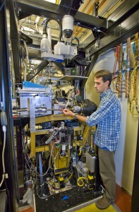
Greg Hura at the ALS SIBYLS beamline, which features two interchangeable end stations, one for macromolecular crystallography and one for small angle X-ray scattering. (Photo by Roy Kaltschmidt)
Greg Hura at the ALS SIBYLS beamline, which features endstations for macromolecular crystallography and small angle X-ray scattering. (Photo by Roy Kaltschmidt)
Hura is the lead author of a paper describing this research in the Proceedings of the National Academy of Sciences (PNAS). The article is titled “DNA conformations in mismatch repair probed in solution by X-ray scattering from gold nanocrystals.” The corresponding author is John Tainer, who holds joint appointments with Berkeley Lab’s Life Sciences Division and the Scripps Research Institute. (See below for complete list of co-authors)

“It is a common belief that DNA is a passive component in protein interactions that involve DNA metabolism, but many proteins actually make use of DNA structural features, such as rigidity and conformation for important biological processes,” Tainer says. “The view of DNA as a passive element is at least in part due to a paucity of robust tools for examining dynamic DNA conformational states during multistep reactions.”

For this study, Tainer, Hura and their colleagues were able to capitalize on the high quality X-ray beams at ALS beamline 12.3.1, also known as SIBYLS, which stands for Structurally Integrated Biology for Life Sciences. Maintained by Berkeley Lab’s Life Sciences Division under the direction of Tainer, the SIBYLS experimental station is optimized for SAXS imaging, which provides global information on the conformations adopted by a population of macromolecules in almost any solution condition.

“Because X-rays scatter predominantly from electrons, the use of  gold nanocrystals provides extremely high contrast relative to organic molecules critical for biology,” Tainer says. “This is important because we can follow specific biological molecules in complex reactions and along pathways to understand how their changes in shape and assembly control the biological outcomes.”

John Tainer, who holds joint appointments with Berkeley Lab and the Scripps Research Institute, directs the experimental stations at the ALS SIBYLS beamline.
The SAXS study at the SIBYLS beamline validated what has been dubbed the “beads-on-a-string” model of DNA repair, in which MutS proteins are the beads and DNA is the string. In solution, the MutS protein will bind to a mismatched DNA site by bending the DNA. ATP enzymes will come in to encircle and excise the error. The MutS then straightens out the bend and continues to proofread the DNA.

“This is the first time we used this technique to look at a protein-mediated process like DNA repair in solution with multiple partners,” Hura says. “We were able to determine some important details about MutS and the MMR system that should be valuable for drug design. We also now know what to look for in cancer-causing mutations of MutS. When we look at mutant versions of MutS we may be able to see that they do not bend the DNA or form the filament to the same extent as the normal version.”

In addition to Hura and Tainer, other co-authors of the PNAS paper were Chi-Lin Tsai, Shelley Claridge, Marc Mendilloc, Jessica Smith, Gareth Williams, Alexander Mastroianni, Paul Alivisatos, Christopher Putnam and Richard Kolodner.
This research was supported by the DOE Office of Science, the National Institutes of Health, and by the Berkeley Laboratory Directed Research and Development program.