Friday, January 31, 2014

Nearly Everyone Uses Piezoelectrics. Be Nice to Know How They Work.

neutron scattering pmn pzt composite
These two neutron scattering images represent the nanoscale structures of single crystals of PMN and PZT. Because the atoms in PMN deviate slightly from their ideal positions, diffuse scattering results in a distinctive "butterfly" shape quite different from
that of PZT, in which the atoms are more regularly spaced.
Credit: NIST
Piezoelectrics—materials that can change mechanical stress to electricity and back again—are everywhere in modern life. Computer hard drives. Loud speakers. Medical ultrasound. Sonar. 
Though piezoelectrics are a widely used technology, there are major gaps in our understanding of how they work. 
Now researchers at the National Institute of Standards and Technology (NIST) and Canada's Simon Fraser University believe they've learned why one of the main classes of these materials, known as relaxors, behaves in distinctly different ways from the rest and exhibit the largest piezoelectric effect. And the discovery comes in the shape of a butterfly.*

"PMN-based relaxors and ferroelectric PZT have been known for decades, but it has been difficult to identify conclusively the origin of the behavioral differences between them because it has been impossible to grow sufficiently large single crystals of PZT," says the NIST Center for Neutron Research (NCNR)'s Peter Gehring. "We've wanted a fundamental explanation of why relaxors exhibit the greatest piezoelectric effect for a long time because this would help guide efforts to optimize this technologically valuable property."The team examined two of the most commonly used piezoelectric compounds—the ferroelectric PZT and the relaxor PMN—which look very similar on a microscopic scale. Both are crystalline materials composed of cube-shaped unit cells (the basic building blocks of all crystals) that contain one lead atom and three oxygen atoms. The essential difference is found at the centers of the cells: in PZT these are randomly occupied by either one zirconium atom or one titanium atom, both of which have the same electric charge, but in PMN one finds either niobium or manganese, which have very different electric charges. The differently charged atoms produce strong electric fields that vary randomly from one unit cell to another in PMN and other relaxors, a situation absent in PZT.
A few years ago, scientists from Simon Fraser University found a way to make crystals of PZT large enough that PZT and PMN crystals could be examined with a single tool for the first time, permitting the first apples-to-apples comparison of relaxors and ferroelectrics. That tool was the NCNR's neutron beams, which revealed new details about where the atoms in the unit cells were located. In PZT, the atoms sat more or less right where they were expected, but in the PMN, their locations deviated from their expected positions—a finding Gehring says could explain the essentials of relaxor behavior.
"The neutron beams scatter off the PMN crystals in a shape that resembles a butterfly," Gehring says. "It gives a characteristic blurriness that reveals the nanoscale structure that exists in PMN—and in all other relaxors studied with this method as well—but does not exist in PZT. It's our belief that this butterfly-shaped scattering might be a characteristic signature of relaxors."
Additional tests the team performed showed that PMN-based relaxors are over 100 percent more sensitive to mechanical stimulation compared to PZT, another first-time measurement. Gehring says he hopes the findings will help materials scientists do more to optimize the behavior of piezoelectrics generally.
*D. Phelan, C. Stock, J.A. Rodriguez-Rivera, S. Chi, J.Leão, X. Long,Y. Xie, A.A. Bokov, Z. Ye, P. Ganesh and P.M. Gehring. Role of random electric fields in relaxors.Proceedings of the National Academy of Sciences, Jan. 21, 2014. DOI:10.1073/pnas.1314780111.

Source: http://www.nist.gov/ncnr/piezo-012914.cfm

How to Make a Better Invisibility Cloak—With Lasers

Images: Clockwise from top left: Karlsruhe Institute of Technology; Karlsruhe Institute of Technology/ Nature Materials; Karlsruhe Institute of Technology; Karlsruhe Institute of Technology/Applied Physics Letters
Made From Scratch: Lasers were used to draw the micrometer-scale structures in these metamaterials. Pictured clockwise from top left are a bichiral photonic crystal [top view], a photonic quasicrystal, a bichiral photonic crystal [oblique view], and a pentamode metamaterial.

Direct laser writing creates metamaterial structures small enough to manipulate visible light


For a century or more, nearly all technological advances have depended on our ability to produce and manipulate the vast variety of materials that nature has given us. Nowhere is that dependence more evident than in the field of electronics. From a smorgasbord of semiconductors, polymers, and metals, we’ve been able to create a dazzling array of circuitry that now underpins pretty much every aspect of modern life.
So now imagine what we could do if we weren’t limited to the materials found in nature. Researchers have long believed that it would someday be possible to produce artificial materials, or “metamaterials,” and that they would bring about some stunning, otherworldly technologies—the sort that have figured in science fiction tales for years. These innovations include invisibility cloaks that could mask the presence of objects or their electromagnetic signatures, “unfeelability cloaks” that could mechanically mask the tactile feel of an object, superlenses that could resolve features too small to be seen with ordinary microscope lenses, and power absorbers that could capture essentially all of the sunlight hitting a solar cell.
To achieve these advances we’ll need better metamaterials, and those are on the way. Metamaterials are made up of “meta-atoms”—small two- or three-dimensional structures made of polymer, dielectric material, or metal. When these structures are arranged in regular, repeating crystals, they can be used to manipulate electromagnetic radiation in new ways. Ultimately, the capabilities of a metamaterial are determined by the size, shape, and quality of these structures. And the technology to fabricate meta-atoms has recently turned a corner.
Over the past few years, research groups around the world have succeeded in developing a way to draw meta-atoms using lasers. The resulting structures can now take on nearly any shape and be stacked in three dimensions in dense, crystal-like arrangements. What’s more, they can be made small enough to exhibit unique mechanical and thermal properties and to alter the flow of light in a range of wavelengths—including the long-inaccessible visible chunk of the spectrum. Thanks to this microscopic fabrication technology, we can finally see a path beyond the materials nature has provided us into entirely new realms that are limited only by our imaginations.
Source: http://spectrum.ieee.org/semiconductors/materials/how-to-make-a-better-invisibility-cloakwith-lasers

Thursday, January 30, 2014

Novel Nanotherapy Breakthrough May Help Reduce Recurrent Heart Attacks and Stroke


Icahn School of Medicine at Mount Sinai designs HDL nanoparticle to deliver statin medication inside inflamed blood vessels to prevent repeat heart attacks and stroke.

Up to 30 percent of heart attack patients suffer a new heart attack because cardiologists are unable to control inflammation inside heart arteries — the process that leads to clots rupturing and causing myocardial infarction or stroke.

But a report in Nature Communications by Icahn School of Medicine at Mount Sinai scientists showcases the development of a new technology that may provide a solution to this high risk of repeat heart attacks — and potentially help save more lives.

An international research team, led by Mount Sinai investigators, designed and tested a high-density lipoprotein (HDL) nanoparticle loaded with a statin drug. In mouse studies, they show this HDL nanotherapy is capable of directly targeting and lowering dangerous inflammation in blood vessels.

Not only could the HDL nanotherapy potentially avert repeat heart attacks, it may also have the power to reduce recurrent strokes caused by clots in brain arteries, says the study's senior investigator, Willem Mulder, PhD, Associate Professor of Radiology in the Translational and Molecular Imaging Institute at the Icahn School of Medicine at Mount Sinai.

"We envision that a safe and effective HDL nanotherapy could substantially lower cardiovascular events during the critical period of vulnerability after a heart attack or stroke," says Dr. Mulder. 

"While we have much more to do to confirm clinical benefit in patients, our study shows how this nanotherapy functions biologically, and how this novel concept could potentially also work in the clinical setting to solve a critical problem," says Dr. Mulder. "This nanotherapy would be the first of its kind."

Inject HDL Statin Nanotherapy Right After Heart Attack and Stroke Treatment

The research team, led by two PhD graduate students as first authors — Raphael Duivenvoorden, MD, and Jun Tang, MS— fashioned the nanoparticle to resemble an HDL cholesterol particle. In fact, the nanoparticle binds on to the same receptors as natural HDL in order to deliver the statin drug.

Oral statin medications used by millions of people today work primarily in the liver to reduce levels of unhealthy lipids, such as low density lipoproteins (LDL), that circulate in the blood. Statins also exert a very weak dampening effect on some inflammatory cells, foremost those called macrophages that hide within plaque in the arterial walls.

It is this anti-inflammatory function that the researchers sought to bolster by designing their HDL nanotherapy.

Inflammation is the main driver of plaque buildup in arteries. Without inflammation, as well as lipid deposition in blood vessels, clots would not form. As inflammation progresses, macrophages secrete enzymes that degrade the walls of blood vessels, leading to a break in the vessel and the formation of clots. These clots can then clog arteries, leading to a heart attack or stroke. 

"Levels of inflammation spike after a heart attack, which is why up to 30 percent of heart attack patients may suffer another heart attack, some while in hospital or just after discharge," says co-author Zahi Fayad, PhD, Professor of Radiology and Director of the Translational and Molecular Imaging Institute at Icahn School of Medicine at Mount Sinai. "This is the vital time to attack this inflammation culprit, which we currently are unable to do clinically right now," says Dr. Fayad. "Even with the most aggressive treatment available, repeat heart attacks do occur."

According to Mount Sinai researchers, the best way to use their HDL nanotherapy is by injection after the clot that produces a heart attack or stroke has been treated. The HDL nanoparticle would deliver the statin directly to macrophages that are driving the inflammatory response. "This could potentially and very rapidly stabilize a dangerous situation," Dr. Mulder says. "In addition, after discharge, patients would continue to use their oral statins to control LDL in their blood."

"Our study also confirms that the HDL nanoparticle is not seen as a foreign invader by the body's immune system and that it has an inherent and natural affinity to target plaque macrophages," says Jun Tang. "Our experiments demonstrated a very rapid reduction in inflammation in mice with advanced plaque buildup."

If the HDL nanotherapy works well in clinical studies, it may be possible to use it in the future as a heart attack prevention tool, according to Dr. Fayad. "If proven to be safe and effective in humans, this would be a critical advance for cardiovascular medicine. We look forward to further testing of our team's novel nanotherapy breakthrough."


A disk brake for molecules

Using centrifugal force to decelerate particles creates new opportunities for chemistry and quantum information processing

Compared with our breath, passenger planes move at a pretty leisurely pace. On the average, nitrogen molecules, for example, travel at a speed of more than 1,700 kilometres per hour at room temperature, or almost one-and-a-half times the speed of sound. This means the particles are much too fast for many experiments, and also some conceivable applications. However, physicists at the Max Planck Institute of Quantum Optics in Garching have now found a rather simple way to slow down polar molecules to about 70 kilometres per hour. They let the molecules of various substances, such as fluoromethane, run up against the centrifugal force on a rotating disk, while being guided by electrodes. The speed of the decelerated molecules corresponds to a temperature of minus 272 degrees Celsius. The new method makes it possible to produce relatively large quantities of cold molecules in a continuous flow, which could be useful, for instance, for targeted chemical reactions of individual particles, or the processing of quantum information.
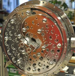
Deceleration in the centrifuge: Molecules lose speed drastically when they are guided against the centrifugal force to the centre of a rotating disk. Electrodes guide the particles to the centre of the centrifuge. The rotating electrodes are likewise designed as disks to ensure mechanical stability. The edges of the disks act like electrostatic guiderails for the molecules. Seen here are the inwardly bent electrodes that are used to guide the particles to the rotation axis. The electrode disks aren’t symmetric with respect to the centre of the disk, so the holes in them balance them out to prevent any imbalance during rotation.Zoom Image
Deceleration in the centrifuge: Molecules lose speed drastically when they are guided against the centrifugal force to... [more]
Chemical reactions are pretty uncontrolled. The reaction partners encounter each other by chance and then collide quite violently, whereupon it is not certain they will do what chemists expect them to do. Bringing them close to each other systematically and at a leisurely pace could favour some transformations that otherwise rarely occur. For this to happen, chemists need slow, and therefore cold, molecules, and they need these in large quantities. Physicists as well rely on cold molecules for many experiments, as well as for new technological applications, such as quantum information processing. For many scientists, especially in low-temperature physics, it should thus be welcome news that researchers working with Sotir Chervenkov and Gerhard Rempe at the Max Planck Institute of Quantum Optics have developed a versatile and efficient brake for polar molecules.
The Garching-based team’s decelerator slows down the particles – in their current experiments, molecules of fluoromethane, trifluoromethane and 3,3,3-trifluoropropine – from about 700 to 70 kilometres per hour. Since the speed of the particles can be expressed in temperature units, this corresponds to reducing the temperature from 100 K to 1 K, or from minus 173 to minus 272 degrees Celsius. “Nitrogen-cooled sources supply molecules at 100 Kelvin, and we also know some good methods for further cooling molecules at 1 Kelvin,” says Sotir Chervenkov. “But there are currently no efficient methods for the range in between, and particularly none that produce a continuous flow of cold molecules.”

Four electrodes guide molecules to the centre of the centrifuge

The Max Planck researchers rely here on an amply known force, but one that has never before been used to slow down molecules: centrifugal force. The molecular brake thus consists of a centrifuge that rotates at up to 43 revolutions per second: a 40-centimetre-in-diameter rotating disk on which the particles are guided from its periphery to its centre.  Four electrodes with alternating polarity spaced one millimetre apart and arranged at the apices of a square serve as guiderails imposing with their electric field a travel direction on the molecules.
The principle of the molecular brake: Four electrodes initially guide polar molecules from the entry of the centrifuge at the lower left along the edge of a rotating disk. They are then guided in a spiral to the centre of the disk. Two static electrodes at the side of the disk are shown in yellow and green, and the electrodes mounted on the disk, in violet and pink.Zoom Image
The principle of the molecular brake: Four electrodes initially guide polar molecules from the entry of the centrifuge... [more]
Two static electrodes gird the disk brake. Through an opening in this double ring, the Max Planck physicists guide the particles into the decelerator. On the disk are likewise mounted, along almost the entire circumference, two electrodes, but not forming closed rings. Rather, the two electrodes bend in a spiral toward the centre across about a quarter of the circular area.
To ensure that there are always four electrostatic guiderails keeping the molecules on track along their deceleration path, a further electrode pair accompanies the particles along the spiral coil. These electrodes are tapered and interface with the static electrode ring at a distance of just 0.2 millimetres, so that it looks as if they branched out of the ring. The molecules are thus moved smoothly onto the curved path, on which they fight against the centrifugal force and drastically lose speed until a further curve in the electrodes in the centre of the disk guides them up and away from the decelerator.

Molecules would have to fly up 2,000 metres against the Earth’s gravitational field

“The deceleration is accomplished in two steps,” explains Martin Zeppenfeld, who originally devised the concept of the molecular brake. “Initially, the molecules slow down when they pass from the laboratory system to the rotating system.” This is comparable to a father running along next to his child on a rotating carousel. He moves with respect to the environment, but for the child, he’s not moving.
“Additionally, the molecules are exposed to the outwardly directed centrifugal force,”  adds Martin Zeppenfeld. “On their way to the centre, the particles must surmount a huge mountain, and are continuously decelerated while doing so, until they finally come almost to a standstill.” For comparison: for the particles to experience the same braking effect in the Earth’s gravitational field, they would have to fly 2,000 meters upward.
Some of the methods currently used to decelerate polar molecules use electrodes not only as guiderails, but also as the actual brake. However, with practicable field strengths, the braking effect remains low, requiring that the particles be sent repeatedly to this electrical potential mountain. This not only results in many particles being lost, but they also don’t leave the decelerator in a continuous flow, but rather in the form of particle pulses, or in other words, in batches.

Centrifuge deceleration is versatile and easy to use

“What is new about our centrifuge deceleration is its continuous operation, the large number of molecules in the resulting beams, its application versatility, and its relative ease of handling,” says Gerhard Rempe, Director at the Max Planck Institute of Quantum Optics. In principle, atoms or neutrons can also be decelerated by a centrifugal force. However, these particles aren’t polar and therefore can’t be guided through the centrifuge using an electric field.
The researchers in Garching now want to further cool the centrifuge-decelerated molecules. They aim to do this using Sisyphus cooling, which they just recently developed, and which is suitable for molecules that are already very cold. Here, an electric field decelerates the optically excited molecules. Through a combination of both methods, the researchers obtain a sufficiently dense flow of extremely cold molecules, allowing them to steer them toward one another to create specific collisions and control their chemical reaction. But the extremely cold molecules could also be accumulated to form clouds that could serve as the register of a quantum computer that is particularly fast for certain arithmetic operations. Thus, the closed cold chain for particles opens up completely new perspectives for chemistry and physics.
Source: http://www.mpg.de/7874908/centrifugal-forces-molecules-brakes

Self-aligning DNA wires for application in nanoelectronics

Since continuous miniaturization in microelectronics is already starting to reach the physical limits, researchers are seeking new methods for device fabrication. One promising candidate is the DNA origami technique in which individual strands of the biomolecule self-assemble into arbitrarily shaped nanostructures. 
The formation of entire circuits, however, requires the controlled positioning of these DNA structures on a surface – something which previously has only been possible using very elaborate techniques. 
Now, researchers at the Helmholtz-Zentrum Dresden-Rossendorf (HZDR) have come up with a simpler strategy which combines DNA origami with self-organized pattern formation. The researchers' method is featured in the scientific journal Nanoscale’s current issue (DOI: 10.1039/C3NR04627C)
Dr. Adrian Keller of the HZDR Institute of Ion Beam Physics and Materials Research describes the new method: “Its beauty lies with the fact that we're allowing nature to simply run its course as soon as we've created the necessary framework.” In the DNA origami technique, the DNA structures self-assemble as long strands of the biomolecule fold into complex, predefined nanoscale shapes by pairing with multiple smaller DNA strands. The physicists used the technique to produce small tubes with lengths of 412 nanometers and diameters of six nanometers. These structures can be used as scaffolds for manufacturing nanoelectronic components like nanowires.
In order to align these nanotubes on the surface, the researchers drew on a principle of self-organization that is actually quite common in nature. Wind may for instance form ordered patterns on a sandy beach. "Similar processes are at work here," explains Keller. "We irradiate the surface onto which we want to place the nanostructures - in our case, the silicon wafers - with ions. This results in the spontaneous appearance of ordered nanopatterns resembling miniature sand dunes. At that point, our job is pretty much done as natural processes are taking over and doing all the work."
Through electrostatic interactions between the charged DNA nanostructures and the charged surface, the nanotubes align themselves in the valleys of the dunes. Says Keller: "This technique works so well that not only do the small tubes follow the wavy patterns, they even replicate occasional pattern defects. Meaning this technique should also allow for production of curved nanocomponents." The maximum degree of alignment the Dresden researchers were able to obtain was at a pattern wavelength of 30 nanometers. "True, we're only looking at a total yield of 70 percent of nanotubes that perfectly follow the pattern," concedes Keller. "But it's still impressive considering the natural process we used."
Because unlike previous approaches, according to Keller, the new technique is quick, cheap, and simple. "Until now, we had to draw on lithographic techniques plus treat the surface with chemicals in order to align the DNA nanostructures. Although this does produce the desired outcome, it nonetheless complicates the processes. Our new technique offers a much simpler alternative." Since aligning the small tubes is based exclusively on electrostatic interaction with the prestructured surface, using this particular method the nanotubes could also be arranged into more complex arrays such as electronic circuits. Keller is convinced that they can be attached to individual transistors, for instance, and connect them electrically: "This way, DNA based nanocomponents could be integrated into technological devices and contribute to further miniaturization."
Developing electronic circuits based on such self-organization principles is the subject of research at the HZDR-coordinated International Helmholtz Research School NanoNet (www.ihrs-nanonet.de). The international Ph.D. program trains junior scientists in molecular electronics as part of DRESDEN-concept – an alliance between the HZDR, the TU Dresden, and several partners from science. The focus of the program is on techniques which functionalize atoms, molecules, and artificial nanostructures to enable information exchange among them and eventually build electronic building blocks like a transistor. The long term vision of this scientific approach is the development of components that spontaneously assemble into electronic circuits.

Publication:
Teshome, B., Facsko, S. & Keller, A. (2014). Topography-controlled alignment of DNA origami nanotubes on nanopatterned surfaces. Nanoscale, 6,1790, DOI: 10.1039/C3NR04627C


Source: http://www.hzdr.de/db/Cms?pNid=99&pOid=40853

Scientists discover long-awaited synthetic particle


Researchers have now created and photographed synthetic magnetic monopoles under laboratory conditions. These observations lay the foundation for the underlying structure of the natural magnetic monopole – the detection of which would be a revolutionary event comparable to the discovery of the electron. The results were recently published in Nature magazine.
Although predicted over 80 years ago, the fundamentally quantum-mechanical configuration of the monopoles has not previously been observed in any physical system. The reported results demonstrate the structure in an ultracold atomic gas.
“Our achievement opens up amazing avenues for quantum research. It feels incredible to have been a part of such a major breakthrough,” says a delighted Dr. Mikko Möttönen from Aalto University, Finland.
Evidence for magnetic monopoles has been sought in sources as diverse as lunar samples and ancient micas. The multibillion-euro LHC particle accelerator at CERN has also been used in the search – but no magnetic monopoles have been convincingly identified. The discovery of the synthetic monopole provides a stronger foundation for these efforts.
“The creation of a synthetic magnetic monopole should provide us with unprecedented insight into aspects of the natural monopole,” says Prof. David S. Hall from Amherst College, USA. “It's not every day that you get to poke and prod the analogue of an elusive fundamental particle under highly controlled conditions in the laboratory.”, he continues.
“Synthesis of the monopole is the starting point for many new breakthroughs in quantum physics research. In the future, we want to get even a more complete correspondence with the natural magnetic monopole.”, says Dr. Möttönen.
A magnetic monopole is a particle just like an electron, but with a magnetic rather than an electric charge. Some 80 years ago Paul A. M. Dirac, one of the founders of quantum physics, discovered a quantum-mechanical structure allowing the existence of magnetic monopoles. Dirac’s original framework has now been experimentally realized for the first time.
Figure caption. Artistic illustration of the synthetic magnetic monopole (Heikka Valja)


Further information

Mikko_Mottonen.jpgMikko Möttönen, docent, Dr. Tech.
Aalto University
Department of Applied Physics and O. V. Lounasmaa Laboratory
QCD Labs
mikko.mottonen*at*aalto.fi
tel. +358 50 594 0950
http://physics.aalto.fi/qcd//
Mikko Möttönen is the leader of the theoretical and computational part of the research. Theoretical ideas and computational modelling was very important for the success of the creation of the monopole. The modelling was carried out using the supercomputers at CSC — IT Center for Science Ltd.

David_S_Hall.jpg
David S. Hall, Professor
Amherst College
Department of Physics
dshall@amherst.edu
tel. +1 413 542 2072
http://www3.amherst.edu/~halllab/
David S. Hall is the leader of the experimental part of the research. The synthetic magnetic monopoles were created in the Physics Laboratories at Amherst College, United States of America.


Funding

This material is based upon work supported by the National Science Foundation,Academy of Finland through its Centres of Excellence Program Computational Nanoscience, and Finnish Doctoral Programme in Computational Sciences.


Background information

Magnetic monopole
“A magnetic monopole is an isolated magnetic pole, magnetic charge, and a point-like source of magnetic field.”
An electron is a point-like particle that carries a so-called elementary electric charge. This means that an electron is an isolated source of an electric field.
Can a magnetic field have a similar point-like source?
Every one of us has likely held two bar magnets and noticed that their ends either attract or repel one another. The ends of the magnet are referred to as poles and every magnet has one end that is a north pole and one that is a south pole. A magnetic north pole attracts a magnetic south pole, but repels another north pole. In general, opposite poles attract, and identical poles repel. In this respect, magnetism is very much like electricity, which exhibits the same attractive and repulsive behavior involving positive and negative electric charges.
When a bar magnet breaks, two smaller bar magnets are created, each with its own north and south pole. You can break each of these smaller magnets in two, and so on, and every resulting magnet has a north pole and a south pole. Even at the atomic level, north and south poles always appear together. One cannot produce in this way a solitary pole, or monopole, that acts as a single point source of the magnetic field.
Are there other ways to find magnetic monopoles?
As yet, not a single natural magnetic monopole has been verifiably observed. This was initially considered to be a problem, because theoretical models that described the post-Big-Bang period predicted that they should be quite common. However, a special model for the expansion of the universe was developed that can explain the extreme rarity of these particles.
According to some theories, the energy content (mass) of a single magnetic monopole is so large that if it were completely used to recharge the battery of an electric car, this vehicle would be able to travel for kilometres with the energy. This explains why magnetic monopoles are probably not likely to occur in a particle accelerator. If the mass of a magnetic monopole really is that large, the energy released from the collision of a negatively and positively charged monopole would be as much as the energy released in the explosion of a kilogram of dynamite!
Dirac monopole
“A Dirac monopole is a point-like source of a possibly artificial magnetic field that forms at the endpoint of a quantum whirlpool.”
In quantum mechanics, an electron is described by a diffuse wave-like object rather than a point-like particle. Paul Dirac was the first person to understand the importance of studying the end points of quantum-mechanical whirlpools within these electron waves. He noticed that when an electron has such a terminating vortex, a magnetic monopole inevitably forms at the end point. A terminating vortex is the defining characteristic of the Dirac monopole.
Dirac also noticed that if the universe contains even a single magnetic monopole, it specifies the smallest possible value for an electric charge. All observed charges must be integer multiples of this minimum value; in other words, charge must be quantized. The existence of a monopole would therefore explain the experimental observation that electric charge is quantized.
Dirac monopoles are generally analyzed in a fairly simple quantum-mechanical model. Magnetic monopoles have since been studied in more general, so-called unified field theories, in which they could exist in the absence of a terminating vortex.
Synthetic magnetic field
“A synthetic magnetic field is an artificial field that leads to particle dynamics equivalent to those of an electric charge in a corresponding natural magnetic field.”
Electrons are not the only physical systems that can exhibit terminating vortices. Thus a Dirac monopole can also appear in other systems, such as the Bose-Einstein condensate. Rather than being related to the natural magnetic field, this monopole can be associated with a synthetic magnetic field. Importantly, the structure of the monopole is identical to that of a Dirac magnetic monopole. This is why the Dirac monopole observed in the synthetic magnetic field is closer to a natural magnetic monopole than any earlier observation.
Spin
“Roughly speaking, spin indicates how fast a particle is spinning around its own axis, and the orientation of that axis.”
Spin is a magnetic property of many particles, including electrons, protons, neutrons, and even many types of atoms. For example, the electron spin is composed of two basis states: up or down. This describes whether the electron is spinning around its axis in a clockwise or counter-clockwise direction.
A particle with a non-zero spin creates a magnetic field around it. However, this is not a monopole field – it is a so-called dipole field with both north and south magnetic poles, just like a bar magnet. Even this smallest of bar magnets cannot be broken into two separate magnetic monopoles.
In fact, bar magnets are composed of countless numbers of small spin dipoles, nearly all of which point in the same direction. Overlapping poles of different sign cancel out the field of each other, and thus the field of an ideal bar magnet looks as if it has magnetic poles only at its ends.
Spins tend to align along an externally applied magnetic field, which is the key to the creation of the synthetic magnetic monopole.
Synthesis of a monopole
“A monopole is created in a Bose–Einstein condensate by using an external magnetic field to guide the spins of the atoms forming the condensate.”
In 2009, Aalto University researchers Ville Pietilä and Mikko Möttönen published theoretical results demonstrating a method to create Dirac monopoles in a Bose–Einstein condensate. The idea involves using external magnetic fields to rotate the atomic spins. A Dirac monopole forms in the condensate as a result of the spin rotation. This method was adopted by the researchers in creating the synthetic magnetic monopole.
The Dirac monopole forms in the artificial magnetic field of the condensate, not in the physical magnetic field which steers the spin degree of freedom. Thus, a natural magnetic monopole is not needed to create the synthetic monopole.
 syntetisointi_HQ-pdfliite.jpg
Caption: Synthesis of a monopole in time, starting from panel and ending with panel c. The arrows show the direction of the physical magnetic field produced in the laboratory. This magnetic field also directs the internal spin degree of freedom of the Bose–Einstein condensate in the direction of the arrows. The end result is that the condensate begins to move as if it were electrically charged and affected by a magnetic monopole in the position marked by the black circle in the image. Click for the full-resolution image.
The Bose–Einstein condensate
“A Bose–Einstein condensate behaves like a single giant atom, even though it can contain millions.”
A Bose–Einstein condensate is sometimes considered to be the fifth state of matter, in addition to solid, liquid, gas, and plasma. In the condensate, the importance and location of individual atoms becomes vague and the system behaves as if it were a single large atom. The first Bose–Einstein condensates were achieved in 1995, and this work received the Nobel Prize in 2001.
“Bose–Einstein condensates provide a window from our world into the quantum wonderland. The more often I peek at it, the more I want to stay there,” says enchanted Dr. Möttönen.
Since Bose–Einstein condensates contain many atoms, photographs of them can be taken using technology that is in part similar to that used in ordinary digital cameras. In addition, the condensates can be forced into the desired shape by means of external magnetic fields and laser beams. These properties make condensates a unique tool for developing new phenomena and quantum technologies. In addition to being used with magnetic monopoles, condensates can simulate the properties of various useful materials to the accuracy of a single atom. One of the daydreams of condensate researchers involves finding a solution for the development of superconducting materials that function at room temperature.
What in the world is quantum physics?
“Quantum physics describes natural phenomena most accurately.”
Quantum physics (also quantum mechanics) is a theory developed over the past 100 years that has been observed to describe the reality in more detail than any other model. It is particularly useful for explaining atomic-level phenomena, which is impossible using classical physics. On the other hand, quantum physics reproduces the same results as classical physics on the large scale.
In quantum mechanics, an electron can take on wave-like properties, sometimes appearing as an extended object rather than a point particle. It is this property of extension, which is shared with Bose-Einstein condensates, that permits the observation of the quantum whirlpools essential to detecting the effect of the magnetic monopole.
Quantum technologies use the laws of quantum physics relieved from classical restrictions to produce practical applications. For example, development of a quantum computer – a potentially super-fast problem solver – is one of the key goals of quantum technologies. A quantum computer would be able to find a solution to certain problems very quickly by using methods that are impossible in the logical framework of a normal computer.
“The laws of quantum physics make it possible to take shortcuts. Among other things, this is the basis of the super-fast speed of a quantum computer,” explains Möttönen.
Future directions
In the future, the research groups will concentrate on more in-depth research into the structure of a synthetic magnetic monopole. They are also interested in the dynamics of monopoles and their interactions with other synthetic particles. One interesting idea involves trying to create a monopole that is not bound to a whirlpool in the same way as is the Dirac monopole. This type of structure could possibly describe a natural magnetic monopole in even more detail.
Source: http://sci.aalto.fi/en/current/news/view/2014-01-29/

Wednesday, January 29, 2014

Energy can be teleported over long distances, say physicists


Squeezed vacuum states could allow long-distance energy teleportation. (Courtesy: iStockphoto/agsandrew)


The ability to teleport energy from one location to another could revolutionise the way quantum devices operate, but only if it can be made to work over practical distances. Now physicists think they know how.


Teleportation is the transfer of an object from one point in the universe to another without travelling through the space in between. It is common practice in many labs around the world. Since the early 90s, physicists have used it to teleport increasingly complex objects starting with photons and more recently with atoms and ions.

But that’s just the beginning. Back in 2010, we looked at the extraordinary work of Masahiro Hotta at Tohoku University in Japan who has worked out that it ought to be possible to teleport energy too. That’s something that could have profound implications for the way quantum devices and machines might be made to work in future.

But energy teleportation has an important limitation–the distance over which it can be sent. The limitations are so severe that it’s hard to see how energy teleportation could help even at the nanoscale. This “strong distance limitation has hampered experimental verification,” says Hotta.

But now he and a couple of mates say they’ve discovered a way round this limitation that allows energy to be teleported over almost any distance. And this new protocol for energy teleportation should allow experimental verification for the first time.

First some background. Energy teleportation relies on the natural quantum variations that occur in a vacuum on the smallest scale. On this scale, a vacuum is far from empty.

Instead, physicists think of it as a maelstrom of virtual quantum particles and antiparticles constantly leaping in and out of existence. That’s OK and does not violate any physical laws as long as the average energy of this vacuum is zero.

It also ensures that regions of space are entangled over these short distances. So what happens in one region immediately influences the region it is entangled with.

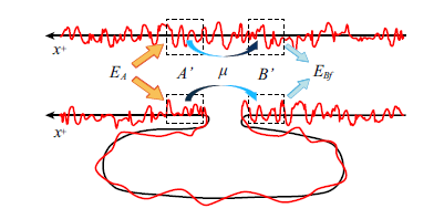
Hotta’s idea is to create a pair of entangled photons and allow one of them to interact with one region of space, thus injecting energy into the vacuum.

It then becomes possible to extract this energy from a nearby, entangled region of space using the other photon. That ensures that any increase in energy in one region is balanced by a decrease in another nearby region.

What limits this process is the distance over which regions of space are entangled, which is not very far, on the order of the Planck scale which is 10^-35 metres. And therein lies the problem.

Now Hotta and pals say they’ve found a way round this using an exotic quantum effect known as a squeezed state, which minimises the quantum noise in a system. They say that preparing the original photons in a squeezed state overcomes the distance limitation.

Instead of relying on entangled regions of space to balance the energy between one point and another, the squeezed state itself does the balancing. And that makes it possible to teleport energy over almost any distance.

Hotta and co say this should make the experimental verification of energy teleportation much easier. If they’re right, we could see the first energy teleportation experiments in the coming months and years.

Ref: arxiv.org/abs/1305.3955 : Quantum Energy Teleportation without Limit of Distance


Source: http://www.technologyreview.com/view/523716/energy-teleportation-overcomes-distance-limit/

Using golden DNA strands to close electric circuits in biosensors

The researchers hope to create diagnostic test which can detect very small amounts of specific DNA
molecules. Photograph: Ken Welch


By letting DNA strands grow together with gold, scientists at Uppsala Berzelii Centre for Neurodiagnostics and Science for Life Laboratory have developed a brand new concept for super sensitive diagnostics of different diseases. The study will be published in the upcoming issue of ACS Nano.

In the new study, the researchers have developed a new method for detecting DNA with an extremely strong signal. The method relies on growth of a DNA strand over a narrow gap between two electrodes in an electric circuit. The strand will only grow if a certain DNA molecule has bound to the surface of one electrode, which makes it possible to build diagnostic tests for detection of that specific DNA molecule.

“We believe that the incredibly strong signal registered when we succeed in growing a golden strand between the electrodes will be possible to turn into a diagnostic test with extreme sensitivity and specificity. Such tests are needed for many diseases where the DNA molecules you are looking for are present only in very small numbers”, says Professor Mats Nilsson, Science for Life Laboratory, who has led the study.

DNA itself does not conduct electricity, but by adding nano particles of gold along the DNA strand, which is then thickened using a gold salt solution, thin gold threads are created within a few minutes which conduct electricity very effectively. When such a strand is created the resistance of the circuit decreases by a factor of a billion.

Camilla Russell, PhD student at Uppsala Berzelii Centre for Neurodiagnostics at Uppsala University and the researcher who has carried out the work, sees great potential in the ‘golden strand’ method: “It should be possible to build very simple test kits using this method, and one test kit can contain many separate sensors to trace a large number of different DNA molecules”, she says.

Fredrik Nikolajeff, Director of Uppsala Berzelii Centre, sees a continued development of the project: “This is a long-term project for developing new sensitive analysis methods which can be used for early diagnostics, for following how diseases develop and to speed up drug development.

We want to continue supporting Camilla’s exciting results through a commercialisation process where we will cooperate with Uppsala University’s business collaboration unit UU Innovation”, he says.

Source: http://www.berzelii.uu.se/uploads/PDF/News/GoldenDNAstrands_in_biosensors_UU_140128.pdf

'Chameleon of the sea' reveals its secrets

Sophisticated biomolecular nanophotonic system underlying the cuttlefish’s color-changing ways


CUTTLEFISH MAY OFFER MODEL FOR BIOINSPIRED HUMAN CAMOUFLAGE AND COLOR-CHANGING PRODUCTS
Scientists at Harvard University and the Marine Biological Laboratory (MBL) hope new understanding of the natural nanoscale photonic device that enables a small marine animal to dynamically change its colors will inspire improved protective camouflage for soldiers on the battlefield.
The cuttlefish, known as the "chameleon of the sea," can rapidly alter both the color and pattern of its skin, helping it blend in with its surroundings and avoid predators. In a paper published January 29 in the Journal of the Royal Society Interface, the Harvard-MBL team reports new details on the sophisticated biomolecular nanophotonic system underlying the cuttlefish’s color-changing ways.
"Nature solved the riddle of adaptive camouflage a long time ago," said Kevin Kit Parker, Tarr Family Professor of Bioengineering and Applied Physics at the Harvard School of Engineering and Applied Sciences (SEAS) and core faculty member at the Wyss Institute for Biologically Inspired Engineering at Harvard. “Now the challenge is to reverse-engineer this system in a cost-efficient, synthetic system that is amenable to mass manufacturing."
In addition to textiles for military camouflage, the findings could also have applications in materials for paints, cosmetics, and consumer electronics.
The cuttlefish (Sepia officinalis) is a cephalopod, like squid and octopuses. Neurally controlled, pigmented organs called chromatophores allow it to change its appearance in response to visual clues, but scientists have had an incomplete understanding of the biological, chemical, and optical functions that make this adaptive coloration possible.
Cuttlefish chromatophores
Left: Cuttlefish chromatophores change from a punctuate to expanded state in response to visual cues. The scale bar measures one millimeter. Right: This illustrated cross-section of the skin shows the layering of three types of chromatophores. Iridophores and leucophores would be positioned beneath the chromatophores. (Images courtesy of Lydia Mathger.)

To regulate its color, the cuttlefish relies on a vertically arranged assembly of three optical components: the leucophore, a near-perfect light scatterer that reflects light uniformly over the entire visible spectrum; the iridophore, a reflector comprising a stack of thin films; and the chromatophore. This layering enables the skin of the animal to selectively absorb or reflect light of different colors, said coauthor Leila F. Deravi, a research associate in bioengineering at Harvard SEAS.
"Chromatophores were previously considered to be pigmentary organs that acted simply as selective color filters,” Deravi said. “But our results suggest that they play a more complex role; they contain luminescent protein nanostructures that enable the cuttlefish to make quick and elaborate changes in its skin pigmentation."
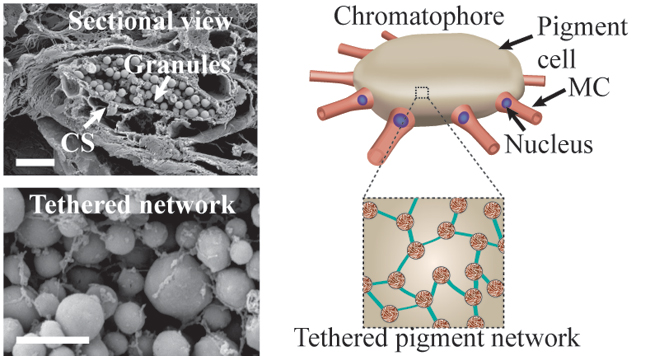
When the cuttlefish actuates its coloration system, each chromatophore expands; the surface area can change as much as 500 percent. The Harvard-MBL team showed that within the chromatophore, tethered pigment granules regulate light through absorbance, reflection, and fluorescence, in effect functioning as nanoscale photonic elements, even as the chromatophore changes in size.
Chromatophore structure
Chromatophores were previously thought to be simply sacs of pigment that acted as filters; scientists have now discovered that nanostructures (labeled here as "granules") within the cells are capable of fluorescing. (Images courtesy of George Bell.)

"The cuttlefish uses an ingenious approach to materials composition and structure, one that we have never employed in our engineered displays," said coauthor Evelyn Hu, Tarr-Coyne Professor of Applied Physics and of Electrical Engineering at SEAS. "It is extremely challenging for us to replicate the mechanisms that the cuttlefish uses. For example, we cannot yet engineer materials that have the elasticity to expand 500 times in surface area. And were we able to do so, the richness of color of the expanded and unexpanded material would be dramatically different—think of stretching and shrinking a balloon. The cuttlefish may have found a way to compensate for this change in richness of color by being an 'active' light emitter (fluorescent), not simply modulating light through passive reflection."
The team also included Roger Hanlon and his colleagues at the Marine Biological Laboratory in Woods Hole, Mass. Hanlon’s lab has examined adaptive coloration in the cuttlefish and other invertebrates for many years.
"Cuttlefish skin is unique for its dynamic patterning and speed of change," Hanlon said. "Deciphering the relative roles of pigments and reflectors in soft, flexible skin is a key step to translating the principles of actuation to materials science and engineering. This collaborative project expanded our breadth of inquiry and uncovered several useful surprises, such as the tether system that connects the individual pigment granules."
For Parker, an Army reservist who completed two tours of duty in Afghanistan, using the cuttlefish to find a biologically inspired design for new types of military camouflage is more than an academic pursuit. He understands first-hand that poor camouflage patterns can cost lives on the battlefield.
"Throughout history, people have dreamed of having an 'invisible suit,'" Parker said. "Nature solved that problem, and now it’s up to us to replicate this genius so, like the cuttlefish, we can avoid our predators."
In addition to Parker, Hu, Hanlon, and Deravi, the coauthors of the Interface paper are: Andrew P. Magyar, a former postdoctoral student in Hu’s group; Sean P. Sheehy, a graduate student in Parker’s group; and George R. R. Bell, Lydia M. Mäthger, Stephen L. Senft, Trevor J. Wardill, and Alan M. Kuzirian, who all work with Hanlon in the Program in Sensory Physiology and Behavior at the Marine Biological Laboratory.
This work was supported in part by the Defense Advanced Research Projects Agency, the Nanoscale Science and Engineering Center at Harvard supported by the National Science Foundation (NSF), the NSF-supported Harvard Materials Research Science and Engineering Center, and the Air Force Office of Scientific Research.

Source: https://www.seas.harvard.edu/news/2014/01/chameleon-of-sea-reveals-its-secrets