Friday, February 28, 2014

A Molecular Ballet under the X-ray Laser

Simulated difference of X-ray diffraction pattern 
from randomly and uniformly aligned molecules. 
The pattern was well reproduced in the experiment. 
Credit: Stephan Stern/CFEL 
Researchers capture snapshots of free molecules by the light of the free electron laser

An international team of researchers has used the world’s most powerful X-ray laser to take snapshots of free molecules. The research team headed by Prof. Jochen Küpper of the Hamburg Center for Free-Electron Laser Science (CFEL) choreographed a kind of molecular ballet in the X-ray beam. With this work, the researchers have cleared important hurdles on the way to X-ray images of individual molecules, as they explain in the scientific journal Physical Review Letters. CFEL is a cooperation of DESY, the University of Hamburg, and the Max Planck Society.

“We have captured the first images of an ensemble of isolated molecules with an X-ray laser,” said DESY scientist Küpper, who is also a professor at the University of Hamburg and a member of the Hamburg Centre for Ultrafast Imaging (CUI) cluster of excellence. “The molecules all posed for the picture in synch.” According to Küpper, this approach opens the way for studies of the ultra-fast dynamics of isolated molecules. There are existing techniques to image single molecules, but none of these is fast enough to catch the ultra-fast motion of molecules.

The conventional way to determine the atomic structure of molecules is to “freeze” them in a crystal and illuminate them with bright X-rays. However, many molecules are extremely difficult to crystallise. In particular, this is a problem with many biomolecules. What’s more, molecules in a crystal can have different properties than molecules in their free form. And molecular dynamics can only be studied to a very limited extent in the crystalline state. Yet exactly this information is in great demand in chemistry, physics, materials research and life sciences. Researchers are therefore working on methods for taking snapshots of individual free molecules.

“The molecules we are investigating are some of the smallest structures in chemistry and biology and consist of just a handful of atoms,” emphasised co-author Dr. Stephan Stern of CFEL. “In order to observe them, you need the most powerful X-ray source on earth, with the shortest exposure time — one ten-trillionth of a second.” The researchers therefore used what is currently the most powerful X-ray laser, the Linac Coherent Light Source (LCLS) at the SLAC National Accelerator Laboratory in California. This free electron laser (FEL) generates short-wave X-ray light by using powerful magnets to send fast electrons from a particle accelerator along a tightly defined slalom course.

In every curve, the fast particles emit flashes of light which add up to an intense laser pulse. These X-ray pulses have such a short wavelength that they can make even atomic dimensions visible. They are also so short and so bright that they can be used to freeze the ultra-fast motion of molecules. But not even this bright light is currently capable of making clear images of single molecules. That’s why the researchers use a trick to study the molecules — they measure how strongly the X-ray light is scattered by the molecules. The molecular structure can be calculated from this diffraction pattern. The more molecules contribute to the diffraction pattern — for example, in a crystal — the clearer it will be.

Instead of a crystal, Küpper’s team illuminated an ensemble of around 100 individual molecules for every single image. However, these molecules must all have the same orientation, so that their diffraction patterns add up and amplify one another. The team took a simple molecule — consisting of a benzene ring with a small nitrile arm of carbon and nitrogen and with two iodine atoms attached, one above and one below. The researchers first sorted the compound — known chemically as di-iodobenzonitrile — using an inhomogeneous electric field, so that only molecules in the same quantum state could wander into the X-ray beam. They then used a special arrangement of lasers to ensure that the particles all took up the same pose for the photo — like the members of a ballet ensemble — so that all of the benzene rings had the two iodine atoms aligned at their top and bottom.

“We sorted the molecules, led them onto the stage, and then got them to pose in synch for the photo,” said Stern. “Then we took the picture with an ultra-short flash of incredible brightness. The exposure time was so short that the superfast motion of the molecules was frozen and we were able to capture a sharp image of the tiny structures.” In this way, the researchers were able to determine that the distance between the two iodine atoms on the benzene ring was 800 picometres (800 billionths of a millimetre), which is in good agreement with the actual value of 700 picometres known from theory.

The experiments thus point the way to the investigation of extremely high-speed molecular dynamics, in particular at the European X-Ray Laser XFEL, which is currently being constructed from the DESY site in Hamburg’s Bahrenfeld district to the neighbouring town of Schenefeld in Schleswig-Holstein. “In future, we will be able to get the molecules to carry out predetermined sequences of movements, like all of them waving their arms”, said Küpper. “We will be able to film these movements by repeating the experiment a large number of times, taking the snapshots at slightly different times and putting the resulting pictures together in a film. Just like a super slow-motion shot in a sports event or a documentary, these films will show the exact sequence of movements of the molecules during chemical reactions with a precision and level of detail that have never before been achieved.”

Researchers from Germany, Denmark, the Netherlands, Sweden and the USA participated in the study.

Source: http://www.desy.de/information__services/press/pressreleases/@@news-view?id=7361

Thursday, February 27, 2014

Need a water filter? Peel a tree branch

A false-color electron microscope image showing E. coli bacteria
(green) trapped over xylem pit membranes (red and blue) in the
sapwood after filtration.
IMAGE COURTESY OF THE RESEARCHERS
If you’ve run out of drinking water during a lakeside camping trip, there’s a simple solution: Break off a branch from the nearest pine tree, peel away the bark, and slowly pour lake water through the stick. The improvised filter should trap any bacteria, producing fresh, uncontaminated water. 

In fact, an MIT team has discovered that this low-tech filtration system can produce up to four liters of drinking water a day — enough to quench the thirst of a typical person. 

In a paper published this week in the journal PLoS ONE, the researchers demonstrate that a small piece of sapwood can filter out more than 99 percent of the bacteria E. coli from water. They say the size of the pores in sapwood — which contains xylem tissue evolved to transport sap up the length of a tree — also allows water through while blocking most types of bacteria. 

Co-author Rohit Karnik, an associate professor of mechanical engineering at MIT, says sapwood is a promising, low-cost, and efficient material for water filtration, particularly for rural communities where more advanced filtration systems are not readily accessible. 

“Today’s filtration membranes have nanoscale pores that are not something you can manufacture in a garage very easily,” Karnik says. “The idea here is that we don’t need to fabricate a membrane, because it’s easily available. You can just take a piece of wood and make a filter out of it.”

The paper’s co-authors include Michael Boutilier and Jongho Lee from MIT, Valerie Chambers from Fletcher-Maynard Academy in Cambridge, Mass., and Varsha Venkatesh from Jericho High School in Jericho, N.Y.

Tapping the flow of sap

There are a number of water-purification technologies on the market today, although many come with drawbacks: Systems that rely on chlorine treatment work well at large scales, but are expensive. Boiling water to remove contaminants requires a great deal of fuel to heat the water. Membrane-based filters, while able to remove microbes, are expensive, require a pump, and can become easily clogged. 

Sapwood may offer a low-cost, small-scale alternative. The wood is comprised of xylem, porous tissue that conducts sap from a tree’s roots to its crown through a system of vessels and pores. Each vessel wall is pockmarked with tiny pores called pit membranes, through which sap can essentially hopscotch, flowing from one vessel to another as it feeds structures along a tree’s length. The pores also limit cavitation, a process by which air bubbles can grow and spread in xylem, eventually killing a tree. The xylem’s tiny pores can trap bubbles, preventing them from spreading in the wood.

“Plants have had to figure out how to filter out bubbles but allow easy flow of sap,” Karnik observes. “It’s the same problem with water filtration where we want to filter out microbes but maintain a high flow rate. So it’s a nice coincidence that the problems are similar.” 

Seeing red

To study sapwood’s water-filtering potential, the researchers collected branches of white pine and stripped off the outer bark. They cut small sections of sapwood measuring about an inch long and half an inch wide, and mounted each in plastic tubing, sealed with epoxy and secured with clamps. 

Before experimenting with contaminated water, the group used water mixed with red ink particles ranging from 70 to 500 nanometers in size. After all the liquid passed through, the researchers sliced the sapwood in half lengthwise, and observed that much of the red dye was contained within the very top layers of the wood, while the filtrate, or filtered water, was clear. This experiment showed that sapwood is naturally able to filter out particles bigger than about 70 nanometers. 

However, in another experiment, the team found that sapwood was unable to separate out 20-nanometer particles from water, suggesting that there is a limit to the size of particles coniferous sapwood can filter. 

Picking the right plant

Finally, the team flowed inactivated, E. coli-contaminated water through the wood filter. When they examined the xylem under a fluorescent microscope, they saw that bacteria had accumulated around pit membranes in the first few millimeters of the wood. Counting the bacterial cells in the filtered water, the researchers found that the sapwood was able to filter out more than 99 percent of E. coli from water. 

Karnik says sapwood likely can filter most types of bacteria, the smallest of which measure about 200 nanometers. However, the filter probably cannot trap most viruses, which are much smaller in size. 

Karnik says his group now plans to evaluate the filtering potential of other types of sapwood. In general, flowering trees have smaller pores than coniferous trees, suggesting that they may be able to filter out even smaller particles. However, vessels in flowering trees tend to be much longer, which may be less practical for designing a compact water filter. 

Designers interested in using sapwood as a filtering material will also have to find ways to keep the wood damp, or to dry it while retaining the xylem function. In other experiments with dried sapwood, Karnik found that water either did not flow through well, or flowed through cracks, but did not filter out contaminants. 

“There’s huge variation between plants,” Karnik says. “There could be much better plants out there that are suitable for this process. Ideally, a filter would be a thin slice of wood you could use for a few days, then throw it away and replace at almost no cost. It’s orders of magnitude cheaper than the high-end membranes on the market today.”

While the pores in sapwood are too big to filter out salts, Saurya Prakash, an assistant professor of mechanical engineering at Ohio State University, says the design could be useful in parts of the world where people collect surface water, which can be polluted with fine dust and particles of decaying plant and animal matter. Most of this detritus, Prakash says, could easily be filtered out by the group’s design. 

“The xylem tissue acts as a natural filter, similar to a manmade membrane,” says Prakash, who was not involved in the research. “The study by the Karnik group shows that use of abundant, naturally occurring materials could pave the way for a new generation of water filters that are potentially low-cost enough to be disposable.”

This research was supported by the James H. Ferry Jr. Fund for Innovation in Research Education.

Source: http://web.mit.edu/newsoffice/2014/need-a-water-filter-peel-a-tree-branch-0226.html

JILA Physicists Discover 'Quantum Droplet' in Semiconductor

Artist's conception of microscopic "quantum droplet" 
discovered by JILA physicists in a gallium-arsenide 
semiconductor excited by an ultrafast red laser pulse. 
Each droplet consists of electrons and holes 
(representing absent electrons) arranged in a 
liquid-like pattern of rings. The surrounding area 
is plasma. The discovery adds to understanding of 
how electrons interact in optoelectronic devices.
Credit: Baxley/JILA
JILA physicists used an ultrafast laser and help from German theorists to discover a new semiconductor quasiparticle—a handful of smaller particles that briefly condense into a liquid-like droplet.

Quasiparticles are composites of smaller particles that can be created inside solid materials and act together in a predictable way. A simple example is the exciton, a pairing, due to electrostatic forces, of an electron and a so-called "hole," a place in the material's energy structure where an electron could be, but isn't.
The new quasiparticle, described in the Feb. 27, 2014, issue of Nature* and featured on the journal's cover, is a microscopic complex of electrons and holes in a new, unpaired arrangement. The researchers call this a "quantum droplet" because it has quantum characteristics such as well-ordered energy levels, but also has some of the characteristics of a liquid. It can have ripples, for example. It differs from a familiar liquid like water because the quantum droplet has a finite size, beyond which the association between electrons and holes disappears.
Although its lifetime is only a fleeting 25 picoseconds (trillionths of a second), the quantum droplet is stable enough for research on how light interacts with specialized forms of matter.
"Electron-hole droplets are known in semiconductors, but they usually contain thousands to millions of electrons and holes," says JILA physicist Steven Cundiff, who studies the properties of cutting-edge lasers and what they reveal about matter. "Here we are talking about droplets with around five electrons and five holes.
"Regarding practical benefits, nobody is going to build a quantum droplet widget. But this does have indirect benefits in terms of improving our understanding of how electrons interact in various situations, including in optoelectronic devices."
The JILA team created the new quasiparticle by exciting a gallium-arsenide semiconductor with an ultrafast red laser emitting about 100 million pulses per second. The pulses initially form excitons, which are known to travel around in semiconductors. As laser pulse intensity increases, more electron-hole pairs are created, with quantum droplets developing when the exciton density reaches a certain level. At that point, the pairing disappears and a few electrons take up positions relative to a given hole. The negatively charged electrons and positively charged holes create a neutral droplet. The droplets are like bubbles held together briefly by pressure from the surrounding plasma.
JILA's experimental data on energy levels of individual droplet rings agreed with theoretical calculations by co-authors at the University of Marburg in Germany. JILA researchers found they could tap into each energy level by tailoring the quantum properties of the laser pulses to match the particle correlations within the droplets. The droplets seem stable enough for future systematic studies on interactions between light and highly correlated states of matter. In addition, quasiparticles, in general, can have exotic properties not found in their constituent parts, and thus, can play a role in controlling the behavior of larger systems and devices.
JILA is a joint institute of the National Institute of Standards and Technology (NIST) and University of Colorado Boulder. Cundiff is a NIST physicist. The JILA research is supported by the National Science Foundation, NIST and the Alexander von Humboldt Foundation.

Source: http://www.nist.gov/pml/div689/droplet-022614.cfm

Wednesday, February 26, 2014

Creating Complex Nanoparticles in One Easy Step


Nanoparticle research is huge.  That is, the study of nanoparticles, very miniscule objects that act as a unit with specific properties, is a very popular area of study.  With implications in many avenues of science, from biomedicine to laser research, the study of how to create nanoparticles with desirable properties is becoming increasingly important.  Maria Benelmekki and researchers in Mukhles Sowwan’s Nanoparticles by Design Unit recently made a breakthrough in synthesizing biomedically relevant nanoparticles.  They published their findings in the journal Nanoscale.
Nanoparticles can be used in medicine for imaging during diagnosis and treatment.  Other applications include targeted drug delivery and wound healing.  However, creating nanoparticles for use in biomedicine presents many challenges.  Currently, nanoparticles are primarily made using chemicals, which is a problem when using them for medical purposes because these chemicals may be harmful to the patient.  Additional issues are that the fabrication process takes several steps, the size of the particles is difficult to control and the particles can only survive in storage for a relatively short amount of time.  Benelmekki and colleagues have created biocompatible ternary nanoparticles, meaning they consist of 3 parts that each exhibit a useful property, and have done it without the use of chemicals.  The new method allows for easy manipulation of the size of the particles to tailor-make them for a variety of uses all in one step.  The researchers have also developed a method that provides better stability for longer storage.
The nanoparticles in the study are made of a core of iron and silver.  These two elements imbue them with two important properties; they are magnetic and can be imaged.  The iron makes them magnetic, allowing researchers to move them around.  The silver is excellent for imaging because excitation of silver creates a larger detection signal than the particle itself, meaning it can be viewed with conventional microscopy or medical imaging devices despite its tiny size.  The third part of the nanoparticles is a silicon shell, which surrounds the iron-silver core.  The silicon is biocompatible, meaning it can go into a patient without creating complications, it prevents the core from being broken down and it can be easily manipulated for use in a variety of biomedical applications.  Additionally, the nanoparticles also have superparamagnetic behavior, meaning they are only magnetic when a magnetic field is applied, so their magnetic property is inducible.
The ability to easily create stable, customizably sized nanoparticles with multiple functionalities, without the use of chemicals, in one step, is an exciting breakthrough.  All of this work was possible because of the extensive expertise of the members of the unit in materials science, and their skills to work in a multidisciplinary environment.  The implications of the work are potentially vast.  Benelmekki says, “The ternary nanoparticles can be used in different applications, such as a contrast agent in MRI, biomagnetic sensors, hyperthermia for cancer treatment and magnetically targeted delivery and transfection.”  Maybe the next time you go in for medical imaging or treatment, nanoparticles designed here at OIST will be part of the treatment.
Source: http://www.oist.jp/news-center/news/2014/2/26/creating-complex-nanoparticles-one-easy-step

Cooking Up New Nanoribbons to Make Better White LEDs

As the world moves away from incandescent light bulbs, light-emitting diodes (LEDs) are growing in popularity. 

They use significantly less energy and have far longer lifetimes than do the traditional incandescent bulbs, which are being phased out, and they don't contain mercury, as do compact fluorescents. LEDs do have a drawback, however. The phosphors that convert the single color produced by an LED into white light tend to produce a cool, bluish glow instead of the warmer, yellower color most people prefer.

Now scientists from Argonne National Laboratory, Oak Ridge National Laboratory, and the University of Georgia are developing new compounds to create nanoribbons that luminesce in different colors, which they can mix together to make a phosphor that provides a more desirable white light that can be tailored for different uses. Using high-brightness x-rays at several different beamlines at the U.S. Department of Energy Office of Science's Advanced Photon Source (APS), they are figuring out how atoms in the materials are arranged. A better understanding of these crystal structures will help them fine-tune their nanoribbons and make more appealing white phosphors based on LEDs.

The materials combine the rare-earth element europium with aluminum oxide to form europium aluminate nanoribbons. Powders of europium oxide (Eu2O3) and aluminum oxide (Al2O3) were mixed together with graphite powder and heated in a vacuum chamber to temperatures above 1000° C. Powders heated to between 1200° and 1400° C formed nanoribbons that luminesced orange. Those heated to 1000° to 1200° glowed green. When more aluminum oxide powder was added and the pressure in the vacuum chamber was raised, from 5 Torr to between 10 and 15 Torr, the resulting nanoribbons shone blue.

It is rare, the researchers say, for a single material to be able to cover the spectrum of visible colors, but it could simplify the creation of phosphors that produce desirable colors when excited by a blue or ultraviolet LED. Scientists would simply have to select the right mix of nanoribbons to get the white light they sought. In a separate experiment, the researchers also produced yellow and red luminescent nanoribbons by adding barium to the europium aluminate.

The researchers first used conventional x-ray powder diffraction to determine the crystal structures of the three types of nanoribbon. They compared the patterns they found to several diffraction databases of known materials and discovered the green nanoribbon was a match for strontium aluminate, so they knew it had a similar crystalline shape.

There was no match for the other two types, though, so they turned to the X-ray Science Division (XSD) beamline 11-BM-B at the Argonne Advanced Photon Source to perform high-resolution synchrotron powder diffraction using a single x-ray energy, which provides initial measurements of the spacings between the atomic planes of the crystal. Using a different scattering technique with a wide range of x-ray energies, XSD beamline 34-1D-E, also at the APS, gave them more detailed information, such as the exact angles between different atomic planes and the presence of crystal defects.

Using XSD beamline 20-BM-B at the APS, they performed x-ray absorption near-edge structure measurements, which focused on europium fluorescence and confirmed that the nanoribbons are indeed europium aluminates, and provided their chemical formulas — EuAl6O10 for the blue and EuAl2O4 for the green and orange. The blue nanoribbons, therefore, are a newly discovered compound. While green photoluminescence at room temperature had been seen before from SrAl2O4, the orange color was new as well.

Specifically pinning down the complete atomic structure of the crystal and tying it to the observed behavior of the material is a complex undertaking. The team now has an average picture of the local spatial arrangement of the elements and the oxygen vacancies — areas where a missing oxygen atom affects the electrical behavior of the material. They are now trying to refine that picture using the ChemMatCARS 15-ID-B advanced crystallography beamline at the APS to give theorists enough information to explain cause and effect and suggest possible ways to tweak the materials' luminescence properties.

The researchers also found a result they had not been looking for. They discovered that when they hit an individual nanoribbon with a microfocused x-ray beam, it not only produced the x-ray diffraction patterns they were using for their measurements, but also generated strong visible light emission. That light appeared not only at the spot where the beam struck, but also at the ends of the ribbons, showing the ribbons were acting as waveguides. That ability to route light of different colors means the nanoribbons may help in the creation of circuits inside optical devices, which use light beams to perform their functions.

Source: http://www.aps.anl.gov/Science/Highlights/Content/APS_SCIENCE_20140224.php

Tuesday, February 25, 2014

Researchers Demonstrate a Quantum Connection Between Light and Mechanics

By using a laser to illuminate an object, physicists in the EPFL’s Laboratory of Photonics and Quantum Measurement have shown that it is possible to control the motion of an object, large enough to be seen with the naked eye, at the level where quantum mechanics dominates. Radiation pressure damping by laser light cools the motion down while the interaction between light and the movement of the oscillator form an intimate connection.
Researchers supported by the Swiss National Science Foundation (SNSF) have demonstrated a microscopic system in which light can be converted into a mechanical oscillation and back. This interaction is so strong that it becomes possible to control the motion of the oscillator at the level where quantum mechanics governs its behavior.
Since the early 20th century, it is known that the movement of objects is ultimately governed by the laws of quantum mechanics, which predict some intriguing phenomena: An object could simultaneously be in two places at the same time, and it should always be moving a little, even at a temperature of absolute zero – the oscillator is then said to be in its quantum ground state. Yet we never experience such behavior in the things we see around us and interact with in daily life.
Quantum strangeness
Indeed, quantum effects can only be observed on very well isolated systems, where the coupling to the surrounding environment is extremely weak. For large objects, the unavoidable coupling quickly washes out the quantum properties, in a process known as decoherence. Until recently, scientists were only able to observe quantum mechanical traits in the motion of tiny systems, such as single atoms or molecules. Now, a team of physicists in the EPFL’s Laboratory of Photonics and Quantum Measurement directed by Tobias Kippenberg has shown that it is possible to control the motion of an object, sufficiently large to be seen with the naked eye, at the level where quantum mechanics dominates. They achieve this by illuminating the object with laser light. The results are published in this week’s edition of Nature magazine*.
A ring of light
The structure is a carefully crafted glass donut on a microchip, with a diameter of 30 micrometres (about one half of a hair’s diameter) which can vibrate at a well-defined frequency. At the same time, it acts as a racetrack for light, which can circle around the circumference of the donut. In turning the bend, the light exerts a little force on the glass surface, an effect called ‘radiation pressure’. Although this force is very small, in these structures it can become appreciable since light circles around the structure up to a million times before being lost. The radiation pressure force can make the ring move, causing it to vibrate like a finger running along the rim of a wineglass. But it can in fact also dampen the vibrations, and thus cool down the oscillatory motion.
Cold, colder, …
Cooling is crucial to reaching the regime of quantum mechanical motion, as this is normally overshadowed by random thermal fluctuations. For this reason, the structure is brought to a temperature of less than one degree above absolute zero. Radiation pressure damping by laser light launched into the donut then cools the motion down by an extra factor 100. The oscillator is cooled so much that it spends a large fraction of the time in its quantum ground state. But even more importantly: The interaction between light and the movement of the oscillator can be made so strong that the two form an intimate connection. A small excitation in the form of a light pulse can fully transform into a small vibration and back again. For the first time, this transformation between light and motion is made to occur within a time that is short enough such that the quantum properties of the original light pulse are not lost in the process through decoherence. By outpacing decoherence, the current results provide a powerful way to control the quantum properties of the oscillator motion, and see the peculiar predictions of quantum mechanics at play in human-made objects.
Source: Swiss National Science Foundation
Image: K-Lab/EPFL

Monday, February 24, 2014

TU Delft achieves maximum light trapping in solar cells

Researchers at TU Delft have opened the way for the realization of the next generation of high-efficiency, cost-effective and ultra-thin crystalline silicon solar cells. They are the first in the world to come very close (99.8%) to the theoretical limit of absorption enhancement (light trapping) in a broad light spectrum range. Their article on light management in ultra-thin silicon is accepted for publication in the journal ACS Photonics.


PhD-student Andrea Ingenito of the Photovoltaic Materials and Devices (PVMD) group at TU Delft has experimentally demonstrated the theoretical limit of the enhancement of light absorption in a thin semiconductor material. Ingenito used wafers of crystalline silicon and experimentally proved the theoretical prediction of the maximal enhancement of light absorption in a semiconductor material. He is the first in the world to come very close (99.8%) to the theoretical limit of absorption enhancement in a broad light spectrum range. An experimental demonstration of this absorption enhancement limit in solar cells has been elusive for the last thirty years.


The PVMD group at TU Delft (Faculty of EEMCS) has world-leading expertise in the design, fabrication, and implementation of light trapping structures in the solar cells. The researchers in the group managed to develop an advanced metal-free light trapping scheme for crystalline silicon wafers. At the front side of the silicon wafers they applied a nano-texture known as black-silicon. At the rear side, they implemented a random pyramidal texture coated with a photonic Dielectric Back Reflector which was designed to exhibit maximal and omni-directional internal reflectance. For wafers thinner than 35 μm the researchers achieved more than 99% (with the photonic reflector) and up to 99.8% (with the silver back reflector) of the theoretical classical absorption limit in the broad light spectrum from 400 to 1200 nm.


Successful implementation of TU Delft’s light trapping scheme in crystalline silicon solar cells requires an adequate surface passivation of the front nano-texture. For this purpose, the researchers at the PVMD group have developed thermal silicon oxide and aluminium oxide passivation layers. TU Delft's trapping scheme together with excellent surface passivation opens the way for the realization of the next generation of high-efficiency, cost-effective and ultra-thin crystalline silicon solar cells.

Ingenito’s article in ACS Photonics was co-authored by supervisors prof. dr. Miro Zeman and dr. Olindo Isabella. The research was carried out in the AdLight project (Advanced light trapping for thin and highly efficient silicon solar cells) funded by Agentschap NL. Solland and ECN have been the partners in the project.

Source: http://www.tudelft.nl/en/current/latest-news/article/detail/tu-delft-realiseert-maximale-light-trapping-in-zonnecellen/

On the road to Mottronics

Epitaxial mismatches in the lattices of nickelate
ultra-thin films can be used to tune the energetic
landscape of Mott materials and thereby control
conductor/insulator transitions.

Researchers at the Advanced Light Source Find Key to Controlling the Electronic and Magnetic Properties of Mott Thin Films


“Mottronics” is a term seemingly destined to become familiar to aficionados of electronic gadgets. Named for the Nobel laureate   Nevill Francis Mott, Mottronics involve materials – mostly metal oxides – that can be induced to transition between electrically conductive and insulating phases. If these phase transitions can be controlled, Mott materials hold great promise for future transistors and memories that feature higher energy efficiencies and faster switching speeds than today’s devices. A team of researchers working at Berkeley Lab’s Advanced Light Source (ALS) have  demonstrated the conducting/insulating phases of ultra-thin films of Mott materials can be controlled by applying an epitaxial strain to the crystal lattice.

“Our work shows how an epitaxial mismatch in the lattice can be used as a knot to tune the energetic landscape of Mott materials and thereby control conductor/insulator transitions,” says Jian Liu, a post-doctoral scholar now with Berkeley Lab’s Materials Sciences Division, who is the lead author on a paper describing this work in the journal Nature Communications. “Through epitaxial strain, we forced nickelate films containing only a few atomic layers into different phases with dramatically different electronic and magnetic properties. While some of these phases are not obtainable in conventional ways, we were able to produce them in a form that is ready for device development.”

The Nature Communications paper is titled “Heterointerface engineered electronic and magnetic phases of NdNiO3 thin films.” The corresponding author is Jak Chakhalian, a professor of physics at the University of Arkansas. Co-authors are Mehdi Kargarian, Mikhail Kareev, Ben Gray, Phil Ryan, Alejandro Cruz, Nadeem Tahir, Yi-De Chuang, Jinghua Guo, James Rondinelli, John Freeland and Gregory Fiete.

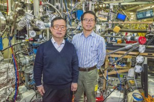
Jinghua Guo (left) and Yi-De Chuang at Beamline 8.0.1 of the Advanced Light Source were part of a team that discovered a key to controlling the electronic and magnetic properties of Mott materials. (Photo by Roy Kaltschmidt)
Nickel-based rare-earth perovskite oxides, or “nickelates,” are considered to be an ideal model for the study of Mott materials because they display strongly correlated electron systems that give rise to unique electronic and magnetic properties. Liu and his co-authors studied thin films of neodymium nickel oxide using ALS beamline 8.0.1, a high flux undulator beamline that produces x-ray beams optimized for the study of nanoscale materials and strongly correlated physics.

“ALS beamline 8.0.1 provides the high photon flux and energy range that are critical when dealing with nanoscale samples,” Liu says. “The state-of-the-art Resonant X-ray Scattering endstation has a high-speed, high-sensitivity CCD camera that makes it feasible to find and track diffraction peaks off a thin film that was only six nanometers thick.”

The transition between the conducting and insulating phases in nickelates is determined by various microscopic interactions, some of which favor the conducting phase, some which favor the insulating phase. The energetic balance of these interactions determines how easily electricity is conducted by electrons moving between the nickel and oxygen ions. By applying enough epitaxial strain to alter the space between these ions, Liu and his colleagues were able to tune this energetic balance and control the conducting/insulating transition. In addition, they   found strain could also be used to control the nickelate’s magnetic properties, again by exploiting the lattice mismatch.

“Magnetism is another hallmark of Mott materials that often goes hand-in-hand with the insulating state and is used to distinguish Mott insulators,” says Liu. “The challenge is that most Mott insulators, including nickelates, are antiferromagnets that macroscopically behave as non-magnetic materials. “At ALS beamline 8.0.1, we were able to directly track the magnetic evolution of our thin films while tuning the metal-to-insulator transition. Our findings give us a better understanding of the physics behind the magnetic properties of these nickelate films and point to potential applications for this magnetism in novel Mottronics devices.”
This research was primarily supported the U.S. Department of Energy’s Office of Science.

Source: http://newscenter.lbl.gov/science-shorts/2014/02/24/on-the-road-to-mottronics/

A Fast and Effective Mechanism to Combat One of the Most Aggressive Cancers


TAU targets drug-resistant ovarian tumors with nanotechnology

Ovarian cancer accounts for more deaths of American women than any other cancer of the female reproductive system. According to the American Cancer Society, one in 72 American women will be diagnosed with ovarian cancer, and one in 100 will ultimately die of the condition.

Now Prof. Dan Peer of Tel Aviv University's Department of Cell Research and Immunology has proposed a new strategy to tackle an aggressive subtype of ovarian cancer using a new nanoscale drug-delivery system designed to target specific cancer cells. He and his team — Keren Cohen and Rafi Emmanuel from Peer's Laboratory of Nanomedicine and Einat Kisin-Finfer and Doron Shabbat, from TAU's Department of Chemistry — have devised a cluster of nanoparticles called gagomers, made of fats and coated with a kind of polysugar. When filled with chemotherapy drugs, these clusters accumulate in tumors, producing dramatically therapeutic benefits.

The objective of Peer's research is two-fold: to provide a specific target for anti-cancer drugs to increase their therapeutic benefits, and to reduce the toxic side effects of anti-cancer therapies. The study was published in February in the journal ACS Nano.

Why chemotherapy fails

According to Prof. Peer, traditional courses of chemotherapy are not an effective line of attack. Chemotherapy's failing lies in the inability of the medicine to be absorbed and maintained within the tumor cell long enough to destroy it. In most cases, the chemotherapy drug is almost immediately ejected by the cancer cell, severely damaging the healthy organs that surround it, leaving the tumor cell intact.

But with their new therapy, Peer and his colleagues saw a 25-fold increase in tumor-accumulated medication and a dramatic dip in toxic accumulation in healthy organs. Tested on laboratory mice, the gagomer mechanism effects a change in drug-resistant tumor cells. Receptors on tumor cells recognize the sugar that encases the gagomer, allowing the binding gagomer to slowly release tiny particles of chemotherapy into the cancerous cell. As more and more drugs accumulate within the tumor cell, the cancer cells begin to die off within 24-48 hours.

"Tumors become resistant very quickly. Following the first, second, and third courses of chemotherapy, the tumors start pumping drugs out of the cells as a survival mechanism," said Prof. Peer. "Most patients with tumor cells beyond the ovaries relapse and ultimately die due to the development of drug resistance. We wanted to create a safe drug-delivery system, which wouldn't harm the body's immune system or organs."

A personal perspective

Prof. Peer chose to tackle ovarian cancer in his research because his mother-in-law passed away at the age of 54 from the disease. "She received all the courses of chemotherapy and survived only a year and a half," he said. "She died from the drug-resistant aggressive tumors.

"At the end of the day, you want to do something natural, simple, and smart. We are committed to try to combine both laboratory and therapeutic arms to create a less toxic, focused drug that combats aggressive drug-resistant cancerous cells," said Prof. Peer. "We hope the concept will be harnessed in the next few years in clinical trials on aggressive tumors," said Prof. Peer.

Source: http://www.aftau.org/site/News2?page=NewsArticle&id=19775

Nanoparticles target anti-inflammatory drugs where needed

Bottom right shows green-labeled neutrophils with
red-labeled nanoparticles inside, which appear yellow

Researchers at the University of Illinois at Chicago have developed a system for precisely delivering anti-inflammatory drugs to immune cells gone out of control, while sparing their well-behaved counterparts. 

Their findings were published online Feb. 23 in Nature Nanotechnology. The system uses nanoparticles made of tiny bits of protein designed to bind to unique receptors found only on neutrophils, a type of immune cell engaged in detrimental acute and chronic inflammatory responses. 

In a normal immune response, neutrophils circulating in the blood respond to signals given off by injured or damaged blood vessels and begin to accumulate at the injury, where they engulf bacteria or debris from injured tissue that might cause infection. In chronic inflammation, neutrophils can pile up at the site of injury, sticking to the blood vessel walls and to each other and contributing to tissue damage. 

Adhesion of neutrophils to blood vessel walls is a major factor in acute lung injury, where it can impair the exchange of gases between the lungs and blood, leading to severe breathing problems. If untreated, the disease has a 50 percent mortality rate in intensive care units.

Corticosteroids and non-steroidal anti-inflammatory drugs used to treat inflammatory diseases are “blunt instruments that affect the whole body and carry some significant side effects,” says Asrar B. Malik, the Schweppe Family Distinguished Professor and head of pharmacology in the UIC College of Medicine, who is lead author of the paper. 

Neutrophils that are stuck to blood vessels or clumped together have unique receptors on their surface that circulating neutrophils lack. Malik and his colleagues designed a nanoparticle to take advantage by embedding it with an anti-inflammatory drug. 

The nanoparticles bind to the receptors, and the neutrophils internalize the nanoparticle. Once inside, the anti-inflammatory drug works to “unzip” the neutrophil and allow it to re-enter the bloodstream. “The nanoparticle is very much like a Trojan horse,” Malik said. “It binds to a receptor found only on these activated, sticky neutrophils, and the cell automatically engulfs whatever binds there. 

Because circulating neutrophils lack these receptors, the system is incredibly precise and targets only those immune cells that are actively contributing to inflammatory disease.” Malik, along with research assistant professor Zhenjia Wang and assistant professor Jaehyung Cho, used intra-vital microscopy to follow nanoparticles in real-time in mice with induced vascular inflammation. 

The nanoparticles were labeled with a fluorescent dye, and could be seen binding to and entering neutrophils clustered together on the inner walls of capillaries, but not binding to freely circulating neutrophils. If the researchers attached a drug called piceatannol, which interferes with cell-cell adhesion, to the nanoparticles, they observed that clusters of neutrophils that took up the particles detached from each other and from the blood vessel wall. 

The cells were in effect neutralized and could no longer contribute to inflammation at the site of an injury. The findings, Malik said, “show that nanoparticles can be used to deliver drugs in a highly targeted, specific fashion to activated immune cells and could be designed to treat a broad range of inflammatory diseases.” Jing Li, postdoctoral research associate in pharmacology, was also a co-author of the study. The research was supported by grants 11SDG7490013 from the American Heart Association, and grants K25HL11157, R01 HL109439 and P01 HL77806 from the National Institutes of Health

Source: http://news.uic.edu/nanoparticles-target-anti-inflammatory-drugs-where-needed#sthash.BLg29h7O.dpuf

Friday, February 21, 2014

Physicists control quantum particles by looking at them

The state of the nuclear spin, visualised by the arrow, after
measurements with varying strengths (light blue is very weak,
dark blue is very strong). For increasing measurement
strength, the state rotates towards the classical up
 state (arrow pointing up). This data is post-selected on one
specific measurement outcome of the electron spin.
For the other outcome, the arrow rotates downwards.
Scientists from the FOM Foundation and TU Delft have manipulated a quantum particle, merely by looking at it in a smart way. By adjusting the strength of their measurement according to earlier measurement outcomes, they managed to steer the particle towards a desired state. The scientists published their results online on 16 February 2014 in Nature Physics.

In two states at once

Quantum mechanics describes the behaviour of microscopic particles, such as atoms and electrons. When we compare it to our observations in everyday life, nature behaves very strangely at the scale of these particles. For instance, an electron can be in two states at the same time.
Schrödinger’s cat in a box, being
both dead and alive at the
same time. When the box is
opened completely, the state
of the cat will be either dead or
alive. By slightly lifting the lid,
it is possible to acquire only a little
bit of information, while maintaining
the fragile quantum state. In this
experiment, the nucleus plays the
role of the cat.
To demonstrate how peculiar this property is, the physicist Erwin Schrödinger proposed a famous thought experiment where the state of a quantum particle is linked to the fate of a cat.
The two are situated in a sealed box. The quantum particle acts as a switch that can either open (switch on) or close (switch off) a small flask of poison. As long as the quantum particle can be simultaneously in two states (on and off), the flask with poison is open and closed, and the cat is both dead and alive at the same time.  
But the weirdness doesn’t end there: as soon as the box is opened to observe the state of the cat, this situation changes. The act of measurement forces the animal to be either dead or alive. This is called the quantum mechanical measurement back-action: the state (of the particle as well as the imaginary cat) is inevitably perturbed by the measurement and collapses to a classical state. In this work, the scientists investigated what happens when the box is only slightly opened. Is it possible to peek at the cat, without destroying the fragile quantum state?

Peeking at Schrödinger’s cat

Instead of a cat, the scientists in the group of FOM workgroup leader prof.dr.ir. Ronald Hanson used a nucleus in diamond. These particles carry an intrinsic property called spin that behaves like a small magnet. The spin of the nucleus can point up (cat alive) or down (cat dead). 

In earlier work the group showed that it is possible to measure the orientation of a single spin, in analogy to fully opening Schrödinger’s box. To partially open the box, the scientists used a trick. Instead of directly measuring the nucleus, they first coupled the state of the nucleus to a nearby electron. They then determined the state of the electron. 
By varying the strength of the coupling between the nucleus and the electron, the scientists could carefully tune the measurement strength. A weaker measurement reveals less information, but also has less back-action. An analysis of the nuclear spin after such a weak measurement showed that the nuclear spin remained in a (slightly altered) superposition of two states. In this way, the scientists verified that the change of the state (induced by the back-action) precisely matched the amount of information that was gained by the measurement. 

Steering by peeking

The scientists realised that it is possible to steer the nuclear spin by applying sequential measurements with varying measurement strength. Since the outcome of a measurement is not known in advance, the researchers implemented a feedback loop in the experiment. They chose the strength of the second measurement depending on the outcome of the first measurement. In this way the scientists could steer the nucleus towards a desired superposition state by only looking at it. 

This result provides new insight in the role of measurements in quantum mechanics. Furthermore the combination of measurements and feedback, as demonstrated here, form an essential building block for the future quantum computer. Finally, these techniques can increase the sensitivity of magnetic field sensors. 
More information
Reference: Manipulating a qubit through the backaction of sequential partial measurements and real-time feedback, M.S. Blok, C. Bonato, M.L. Markham, D.J. Twitchen, V.V. Dobrovitski, R. Hanson, Nature Physics. DOI: 10.1038/nphys2881.
Source: http://home.tudelft.nl/en/current/latest-news/article/detail/sturen-door-gluren-fysici-bedwingen-quantumdeeltjes-door-ze-te-bekijken/

Thursday, February 20, 2014

ORNL microscopy system delivers real-time view of battery electrochemistry

A new in situ transmission electron microscopy technique
enabled  ORNL researchers to image the snowflake-like
growth of the  solid electrolyte interphase from a working
battery electrode. 
Using a new microscopy method, researchers at the Department of Energy’s Oak Ridge National Laboratory can image and measure electrochemical processes in batteries in real time and at nanoscale resolution.
Scientists at ORNL used a miniature electrochemical liquid cell that is placed in a transmission electron microscope to study an enigmatic phenomenon in lithium-ion batteries called the solid electrolyte interphase, or SEI, as described in a study published in Chemical Communications.
The SEI is a nanometer-scale film that forms on a battery’s negative electrode due to electrolyte decomposition. Scientists agree that the SEI’s formation and stability play key roles in controlling battery functionality. But after three decades of research in the battery field, details of the SEI’s dynamics, structure and chemistry during electrochemical cycling are still debated, stemming from inherent difficulties in studying battery electrode materials in their native liquid environment.
 “We’ve used this novel in situ method to understand the dynamics of how this layer forms and evolves during battery operation,” said Raymond Unocic, ORNL R&D staff scientist.
Battery researchers typically study the structure and chemistry of the SEI through “post-mortem” methods, in which a cycled battery is disassembled, dried and then analyzed through a number of characterization methods.
“This is problematic because of the air and moisture sensitivity of the SEI, and the ease by which environmental exposure can modify its structure and chemistry.” Unocic said.
The ORNL researchers formed a miniature electrochemical cell by enclosing battery electrolyte between two silicon microchip devices that contain microfabricated electrodes and silicon nitride viewing membranes. The transparent “windows” seal the highly volatile battery electrolyte from the microscope’s vacuum environment and allow the electron beam to pass through the liquid, which facilitates imaging of the electrochemical reaction products as they form.  
To reproduce a battery charging cycle, the researchers applied a potential at the working electrode and monitored the resulting changes in current. The most striking result, said the researchers, was capturing an unprecedented view of SEI evolution during potential cycling. The technique is able to image the formation of tiny crystalline particles only one billionth of a meter in size.
“As we start to sweep the potential, we didn’t initially observe anything,” said lead author Robert Sacci, a postdoctoral research fellow with ORNL’s FIRST Energy Frontier Research Center. “Then we started seeing shadows -- presumably polymeric SEI -- forming into a dendritic pattern. It looks like a snowflake forming from the electrode.” Watch a video of the process at http://www.youtube.com/watch?v=OHrlFNB-Q9Y.
The researchers plan to build on this initial proof-of-principle study to better understand the factors behind the SEI’s formation, which could ultimately help improve battery performance, capacity, and safety at the device level.
“Tailoring the SEI’s structure and chemistry to maximize battery capabilities appears to be a delicate balancing act,” Unocic said. “When you cycle a real battery, the interphase structure can form, break, and reform again, depending on how thick the layer grows, so we need to look at improving its structural stability. But at the same time, we have to think about making the interphase more efficient for lithium ion transport. This study brings us one step closer to understanding SEI formation and growth.”
Next steps for the researchers include applying their technique to study different types of battery electrodes and electrolytes and other energy storage systems including fuel cells and supercapacitors.
Coauthors are ORNL’s Raymond Unocic, Robert Sacci, Nancy Dudney and Karren More; and Pacific Northwest National Laboratory’s Lucas Parent, Ilke Arslan, Nigel Browning. The study is published as “Direct Visualization of Initial SEI Morphology and Growth Kinetics During Lithium Deposition by in situ Electrochemical Transmission Electron Microscopy.”
The research was supported by the DOE’s Office of Energy Efficiency and Renewable Energy, Vehicle Technologies Program, by the Fluid Interface Reactions Structures and Transport (FIRST) Center, an Energy Frontier Research Center funded by the Office of Basic Energy Sciences, and as part of a user proposal at ORNL’s Center for Nanophase Materials Sciences. Parts of the work were supported by the laboratory directed research and development program at Pacific Northwest National Laboratory and the Joint Center for Energy Storage Research, an Energy Innovation Hub funded by BES-DOE.
Part of this research was conducted at the Center for Nanophase Materials Sciences, which is sponsored at ORNL by the Scientific User Facilities Division in DOE’s Office of Basic Energy Sciences. CNMS is one of the five DOE Nanoscale Science Research Centers supported by the DOE Office of Science, premier national user facilities for interdisciplinary research at the nanoscale. Together the NSRCs comprise a suite of complementary facilities that provide researchers with state-of-the-art capabilities to fabricate, process, characterize and model nanoscale materials, and constitute the largest infrastructure investment of the National Nanotechnology Initiative. The NSRCs are located at DOE's Argonne, Brookhaven, Lawrence Berkeley, Oak Ridge and Sandia and Los Alamos national laboratories. For more information about the DOE NSRCs, please visit http://science.energy.gov/bes/suf/user-facilities/nanoscale-science-research-centers/.
UT-Battelle manages ORNL for the Department of Energy’s Office of Science. DOE’s Office of Science is the single largest supporter of basic research in the physical sciences in the United States, and is working to address some of the most pressing challenges of the time. For more information, please visit science.energy.gov.
Source: http://www.ornl.gov/ornl/news/news-releases/2014/ornl-microscopy-system-delivers-real-time-view-of-battery-electrochemistry